 Movie
Movie Controller

[English] 日本語
 Yorodumi
- PDB-6ozw: Crystal structure of the 65-kilodalton amino-terminal fragment of... -
+ Open data

- Basic information
| Entry | Database: PDB / ID: 6ozw | ||||||
|---|---|---|---|---|---|---|---|
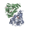
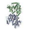
| Title | Crystal structure of the 65-kilodalton amino-terminal fragment of DNA topoisomerase I from Streptococcus mutans | ||||||
 Components Components | DNA topoisomerase 1 | ||||||
 Keywords Keywords | ISOMERASE / DNA BINDING PROTEIN / topoisomerase / supercoiled | ||||||
| Function / homology |  Function and homology information Function and homology informationDNA topoisomerase / DNA topoisomerase type I (single strand cut, ATP-independent) activity / DNA topological change / chromosome / DNA binding / metal ion binding Similarity search - Function | ||||||
| Biological species |  Streptococcus mutans serotype c (bacteria) Streptococcus mutans serotype c (bacteria) | ||||||
| Method |  X-RAY DIFFRACTION / SYNCHROTRON / MOLECULAR REPLACEMENT / molecular replacement / Resolution: 2.063 Å X-RAY DIFFRACTION / SYNCHROTRON / MOLECULAR REPLACEMENT / molecular replacement / Resolution: 2.063 Å | ||||||
 Authors Authors | Jones, J.A. / Hevener, K.E. | ||||||
| Funding support |  United States, 1items United States, 1items
| ||||||
 Citation Citation | Journal: Biochem.Biophys.Res.Commun. / Year: 2019 Title: Crystal structure of the 65-kilodalton amino-terminal fragment of DNA topoisomerase I from the gram-positive model organism Streptococcus mutans. Authors: Jones, J.A. / Hevener, K.E. | ||||||
| History |
|
- Structure visualization
| Structure viewer | Molecule: MolmilJmol/JSmol |
|---|
- Downloads & links
-Download
| PDBx/mmCIF format | 6ozw.cif.gz | 133.4 KB | Display | PDBx/mmCIF format |
|---|---|---|---|---|
| PDB format | pdb6ozw.ent.gz | 99.9 KB | Display | PDB format |
| PDBx/mmJSON format | 6ozw.json.gz | Tree view | PDBx/mmJSON format | |
| Others |  Other downloads Other downloads |
-Validation report
| Arichive directory | https://data.pdbj.org/pub/pdb/validation_reports/oz/6ozwftp://data.pdbj.org/pub/pdb/validation_reports/oz/6ozw | HTTPS FTP |
|---|
-Related structure data
| Related structure data |  1eclS S: Starting model for refinement |
|---|---|
| Similar structure data |


-Links
 PDBj
PDBj


- Assembly
| Deposited unit | 
| ||||||||
|---|---|---|---|---|---|---|---|---|---|
| 1 |
| ||||||||
| Unit cell |
|
-Components
| #1: Protein | Mass: 67783.391 Da / Num. of mol.: 1 / Fragment: residues 1-575 Source method: isolated from a genetically manipulated source Source: (gene. exp.) Streptococcus mutans serotype c (strain ATCC 700610 / UA159) (bacteria)Strain: ATCC 700610 / UA159 / Gene: topA, SMU_1002 / Plasmid: pET-15b / Production host: References: UniProt: Q8DUD3, DNA topoisomerase (ATP-hydrolysing) | ||||
|---|---|---|---|---|---|
| #2: Chemical | ChemComp-MG /   Mass: 24.305 Da / Num. of mol.: 6 / Source method: obtained synthetically / Formula: Mg Mass: 24.305 Da / Num. of mol.: 6 / Source method: obtained synthetically / Formula: Mg#3: Chemical | ChemComp-FMT /   Mass: 46.025 Da / Num. of mol.: 14 / Source method: obtained synthetically / Formula: CH2O2 Mass: 46.025 Da / Num. of mol.: 14 / Source method: obtained synthetically / Formula: CH2O2#4: Water | ChemComp-HOH / |  Mass: 18.015 Da / Num. of mol.: 313 / Source method: isolated from a natural source / Formula: H2O Mass: 18.015 Da / Num. of mol.: 313 / Source method: isolated from a natural source / Formula: H2O |
-Experimental details
-Experiment
| Experiment | Method: X-RAY DIFFRACTION / Number of used crystals: 1 |
|---|
- Sample preparation
| Crystal | Density Matthews: 2.57 Å3/Da / Density % sol: 52.17 % |
|---|---|
| Crystal grow | Temperature: 277.15 K / Method: vapor diffusion, hanging drop Details: 4.6 mg mL-1 in a 3uL:3uL 1:1 ratio protein to condition, crystallization condition 0.2 M magnesium formate dihydrate, 25% w/v PEG 3350 at 277.15 K |
-Data collection
| Diffraction | Mean temperature: 100 K / Serial crystal experiment: N | |||||||||||||||||||||||||||||||||||||||||||||||||||||||||||||||||||||||||||||||||||||||||||||||||||||||||||||||||||||||||||||||||||||||||||||||||||||||||||||||||||||||||||||||||||||||||||||
|---|---|---|---|---|---|---|---|---|---|---|---|---|---|---|---|---|---|---|---|---|---|---|---|---|---|---|---|---|---|---|---|---|---|---|---|---|---|---|---|---|---|---|---|---|---|---|---|---|---|---|---|---|---|---|---|---|---|---|---|---|---|---|---|---|---|---|---|---|---|---|---|---|---|---|---|---|---|---|---|---|---|---|---|---|---|---|---|---|---|---|---|---|---|---|---|---|---|---|---|---|---|---|---|---|---|---|---|---|---|---|---|---|---|---|---|---|---|---|---|---|---|---|---|---|---|---|---|---|---|---|---|---|---|---|---|---|---|---|---|---|---|---|---|---|---|---|---|---|---|---|---|---|---|---|---|---|---|---|---|---|---|---|---|---|---|---|---|---|---|---|---|---|---|---|---|---|---|---|---|---|---|---|---|---|---|---|---|---|---|---|
| Diffraction source | Source: SYNCHROTRON / Site: APS / Beamline: 23-ID-D / Wavelength: 1.0332 Å | |||||||||||||||||||||||||||||||||||||||||||||||||||||||||||||||||||||||||||||||||||||||||||||||||||||||||||||||||||||||||||||||||||||||||||||||||||||||||||||||||||||||||||||||||||||||||||||
| Detector | Type: DECTRIS PILATUS3 6M / Detector: PIXEL / Date: Jun 19, 2018 | |||||||||||||||||||||||||||||||||||||||||||||||||||||||||||||||||||||||||||||||||||||||||||||||||||||||||||||||||||||||||||||||||||||||||||||||||||||||||||||||||||||||||||||||||||||||||||||
| Radiation | Protocol: SINGLE WAVELENGTH / Monochromatic (M) / Laue (L): M / Scattering type: x-ray | |||||||||||||||||||||||||||||||||||||||||||||||||||||||||||||||||||||||||||||||||||||||||||||||||||||||||||||||||||||||||||||||||||||||||||||||||||||||||||||||||||||||||||||||||||||||||||||
| Radiation wavelength | Wavelength: 1.0332 Å / Relative weight: 1 | |||||||||||||||||||||||||||||||||||||||||||||||||||||||||||||||||||||||||||||||||||||||||||||||||||||||||||||||||||||||||||||||||||||||||||||||||||||||||||||||||||||||||||||||||||||||||||||
| Reflection | Resolution: 2.06→50 Å / Num. obs: 39033 / % possible obs: 97.6 % / Redundancy: 6.5 % / Biso Wilson estimate: 24.78 Å2 / Rmerge(I) obs: 0.077 / Rpim(I) all: 0.032 / Rrim(I) all: 0.084 / Χ2: 0.941 / Net I/σ(I): 7.3 / Num. measured all: 254919 | |||||||||||||||||||||||||||||||||||||||||||||||||||||||||||||||||||||||||||||||||||||||||||||||||||||||||||||||||||||||||||||||||||||||||||||||||||||||||||||||||||||||||||||||||||||||||||||
| Reflection shell | Diffraction-ID: 1
|
-Phasing
| Phasing | Method: molecular replacement |
|---|
- Processing
| Software |
| |||||||||||||||||||||||||||||||||||||||||||||||||||||||||||||||||||||||||||||||||||||||||||
|---|---|---|---|---|---|---|---|---|---|---|---|---|---|---|---|---|---|---|---|---|---|---|---|---|---|---|---|---|---|---|---|---|---|---|---|---|---|---|---|---|---|---|---|---|---|---|---|---|---|---|---|---|---|---|---|---|---|---|---|---|---|---|---|---|---|---|---|---|---|---|---|---|---|---|---|---|---|---|---|---|---|---|---|---|---|---|---|---|---|---|---|---|
| Refinement | Method to determine structure: MOLECULAR REPLACEMENT Starting model: 1ECL Resolution: 2.063→49.183 Å / SU ML: 0.3 / Cross valid method: THROUGHOUT / σ(F): 1.37 / Phase error: 30.04
| |||||||||||||||||||||||||||||||||||||||||||||||||||||||||||||||||||||||||||||||||||||||||||
| Solvent computation | Shrinkage radii: 0.9 Å / VDW probe radii: 1.11 Å | |||||||||||||||||||||||||||||||||||||||||||||||||||||||||||||||||||||||||||||||||||||||||||
| Displacement parameters | Biso max: 92.86 Å2 / Biso mean: 33.1822 Å2 / Biso min: 9.72 Å2 | |||||||||||||||||||||||||||||||||||||||||||||||||||||||||||||||||||||||||||||||||||||||||||
| Refinement step | Cycle: final / Resolution: 2.063→49.183 Å
| |||||||||||||||||||||||||||||||||||||||||||||||||||||||||||||||||||||||||||||||||||||||||||
| Refine LS restraints |
| |||||||||||||||||||||||||||||||||||||||||||||||||||||||||||||||||||||||||||||||||||||||||||
| LS refinement shell | Refine-ID: X-RAY DIFFRACTION / Rfactor Rfree error: 0 / Total num. of bins used: 12
|
