 Movie
Movie Controller

[English] 日本語
 Yorodumi
- PDB-2yf9: STRUCTURAL AND FUNCTIONAL INSIGHTS OF DR2231 PROTEIN, THE MAZG-LI... -
+ Open data

- Basic information
| Entry | Database: PDB / ID: 2yf9 | |||||||||
|---|---|---|---|---|---|---|---|---|---|---|
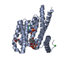
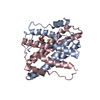
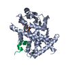
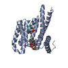
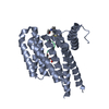
| Title | STRUCTURAL AND FUNCTIONAL INSIGHTS OF DR2231 PROTEIN, THE MAZG-LIKE NUCLEOSIDE TRIPHOSPHATE PYROPHOSPHOHYDROLASE FROM DEINOCOCCUS RADIODURANS, NATIVE FORM | |||||||||
 Components Components | MAZG-LIKE NUCLEOSIDE TRIPHOSPHATE PYROPHOSPHOHYDROLASE | |||||||||
 Keywords Keywords | HYDROLASE / DIMERIC DUTPASE | |||||||||
| Function / homology |  Function and homology information Function and homology information | |||||||||
| Biological species |  DEINOCOCCUS RADIODURANS (radioresistant) DEINOCOCCUS RADIODURANS (radioresistant) | |||||||||
| Method |  X-RAY DIFFRACTION / SYNCHROTRON / MOLECULAR REPLACEMENT / Resolution: 1.899 Å X-RAY DIFFRACTION / SYNCHROTRON / MOLECULAR REPLACEMENT / Resolution: 1.899 Å | |||||||||
 Authors Authors | Goncalves, A.M.D. / De Sanctis, D. / Mcsweeney, S.M. | |||||||||
 Citation Citation | Journal: J.Biol.Chem. / Year: 2011 Title: Structural and Functional Insights Into Dr2231 Protein, the Mazg-Like Nucleoside Triphosphate Pyrophosphohydrolase from Deinococcus Radiodurans. Authors: Goncalves, A.M.D. / Desanctis, D. / Mcsweeney, S.M. | |||||||||
| History |
|
- Structure visualization
| Structure viewer | Molecule: MolmilJmol/JSmol |
|---|
- Downloads & links
-Download
| PDBx/mmCIF format | 2yf9.cif.gz | 92.6 KB | Display | PDBx/mmCIF format |
|---|---|---|---|---|
| PDB format | pdb2yf9.ent.gz | 72.6 KB | Display | PDB format |
| PDBx/mmJSON format | 2yf9.json.gz | Tree view | PDBx/mmJSON format | |
| Others |  Other downloads Other downloads |
-Validation report
| Arichive directory | https://data.pdbj.org/pub/pdb/validation_reports/yf/2yf9ftp://data.pdbj.org/pub/pdb/validation_reports/yf/2yf9 | HTTPS FTP |
|---|
-Related structure data
| Related structure data |  2yeuSC  2yf3C  2yf4C  2yfcC 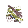 2yfdC C: citing same article ( S: Starting model for refinement |
|---|---|
| Similar structure data |


-Links
 PDBj
PDBj- Assembly
| Deposited unit | 
| ||||||||||||
|---|---|---|---|---|---|---|---|---|---|---|---|---|---|
| 1 | 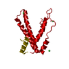
| ||||||||||||
| Unit cell |
| ||||||||||||
| Components on special symmetry positions |
|
-Components
| #1: Protein | Mass: 16708.691 Da / Num. of mol.: 1 Source method: isolated from a genetically manipulated source Source: (gene. exp.) DEINOCOCCUS RADIODURANS (radioresistant)Strain: R1 / Plasmid: PET151/D-TOPO / Production host: References: UniProt: Q9RS96, nucleoside-triphosphate diphosphatase | ||
|---|---|---|---|
| #2: Chemical | ChemComp-CL /   Mass: 35.453 Da / Num. of mol.: 4 / Source method: obtained synthetically / Formula: Cl Mass: 35.453 Da / Num. of mol.: 4 / Source method: obtained synthetically / Formula: Cl#3: Water | ChemComp-HOH / |  Mass: 18.015 Da / Num. of mol.: 142 / Source method: isolated from a natural source / Formula: H2O Mass: 18.015 Da / Num. of mol.: 142 / Source method: isolated from a natural source / Formula: H2O |
-Experimental details
-Experiment
| Experiment | Method: X-RAY DIFFRACTION / Number of used crystals: 1 |
|---|
- Sample preparation
| Crystal | Density Matthews: 2.86 Å3/Da / Density % sol: 57 % / Description: NONE |
|---|---|
| Crystal grow | pH: 7 Details: 1.0 M LITHIUM CHLORIDE, 0.1 M CITRIC ACID, 5-10%(W/V) PEG 6000., pH 7 |
-Data collection
| Diffraction | Mean temperature: 100 K |
|---|---|
| Diffraction source | Source: SYNCHROTRON / Site: ESRF  / Beamline: ID29 / Wavelength: 0.97627 / Beamline: ID29 / Wavelength: 0.97627 |
| Detector | Type: ADSC QUANTUM 315r / Detector: CCD / Date: Feb 22, 2010 / Details: MIRROR |
| Radiation | Monochromator: SI(111) / Protocol: SINGLE WAVELENGTH / Monochromatic (M) / Laue (L): M / Scattering type: x-ray |
| Radiation wavelength | Wavelength: 0.97627 Å / Relative weight: 1 |
| Reflection | Resolution: 1.9→45.68 Å / Num. obs: 16383 / % possible obs: 99.7 % / Observed criterion σ(I): 2 / Redundancy: 9.8 % / Biso Wilson estimate: 23.21 Å2 / Rmerge(I) obs: 0.1 / Net I/σ(I): 14.7 |
| Reflection shell | Resolution: 1.9→2 Å / Redundancy: 9.5 % / Rmerge(I) obs: 0.46 / Mean I/σ(I) obs: 4.2 / % possible all: 98.2 |
- Processing
| Software |
| |||||||||||||||||||||||||||||||||||||||||||||||||
|---|---|---|---|---|---|---|---|---|---|---|---|---|---|---|---|---|---|---|---|---|---|---|---|---|---|---|---|---|---|---|---|---|---|---|---|---|---|---|---|---|---|---|---|---|---|---|---|---|---|---|
| Refinement | Method to determine structure: MOLECULAR REPLACEMENT Starting model: PDB ENTRY 2YEU Resolution: 1.899→45.683 Å / SU ML: 0.22 / σ(F): 1.37 / Phase error: 17.83 / Stereochemistry target values: ML
| |||||||||||||||||||||||||||||||||||||||||||||||||
| Solvent computation | Shrinkage radii: 0.9 Å / VDW probe radii: 1.11 Å / Solvent model: FLAT BULK SOLVENT MODEL / Bsol: 56.991 Å2 / ksol: 0.359 e/Å3 | |||||||||||||||||||||||||||||||||||||||||||||||||
| Displacement parameters | Biso mean: 25.38 Å2
| |||||||||||||||||||||||||||||||||||||||||||||||||
| Refinement step | Cycle: LAST / Resolution: 1.899→45.683 Å
| |||||||||||||||||||||||||||||||||||||||||||||||||
| Refine LS restraints |
| |||||||||||||||||||||||||||||||||||||||||||||||||
| LS refinement shell |
| |||||||||||||||||||||||||||||||||||||||||||||||||
| Refinement TLS params. | Method: refined / Origin x: 28.4473 Å / Origin y: 13.7702 Å / Origin z: 12.1328 Å
| |||||||||||||||||||||||||||||||||||||||||||||||||
| Refinement TLS group | Selection details: CHAIN A |
