 Movie
Movie Controller

[English] 日本語
 Yorodumi
- PDB-2yby: Structure of domains 6 and 7 of the mouse complement regulator Fa... -
+ Open data

- Basic information
| Entry | Database: PDB / ID: 2yby | |||||||||
|---|---|---|---|---|---|---|---|---|---|---|
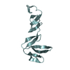
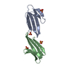
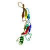
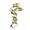
| Title | Structure of domains 6 and 7 of the mouse complement regulator Factor H | |||||||||
 Components Components | COMPLEMENT FACTOR H | |||||||||
 Keywords Keywords | IMMUNE SYSTEM / COMPLEMENT REGULATION / INNATE IMMUNITY / INFECTION | |||||||||
| Function / homology |  Function and homology information Function and homology informationmitochondrial gene expression / photoreceptor cell differentiation / immune complex clearance / retinal rod cell development / activation of membrane attack complex / regulation of complement activation, alternative pathway / organ or tissue specific immune response / vascular associated smooth muscle cell differentiation / Regulation of Complement cascade / regulation of complement-dependent cytotoxicity ...mitochondrial gene expression / photoreceptor cell differentiation / immune complex clearance / retinal rod cell development / activation of membrane attack complex / regulation of complement activation, alternative pathway / organ or tissue specific immune response / vascular associated smooth muscle cell differentiation / Regulation of Complement cascade / regulation of complement-dependent cytotoxicity / monocyte aggregation / retinal pigment epithelium development / mitochondrial DNA metabolic process / regulation of complement activation / inflammatory response to wounding / complement component C3b binding / symbiont cell surface / glomerulus development / neutrophil homeostasis / serine-type endopeptidase complex / heparan sulfate proteoglycan binding / complement activation / complement activation, alternative pathway / cartilage development / neuromuscular process / response to dietary excess / immunoglobulin mediated immune response / ATP metabolic process / visual perception / response to cytokine / determination of adult lifespan / kidney development / mitochondrion organization / bone development / platelet aggregation / heparin binding / cellular response to lipopolysaccharide / retina development in camera-type eye / gene expression / angiogenesis / immune response / inflammatory response / external side of plasma membrane / axon / neuronal cell body / cell surface / mitochondrion / proteolysis / : / extracellular region / identical protein binding Similarity search - Function | |||||||||
| Biological species |  | |||||||||
| Method |  X-RAY DIFFRACTION / SYNCHROTRON / MOLECULAR REPLACEMENT / Resolution: 1.58 Å X-RAY DIFFRACTION / SYNCHROTRON / MOLECULAR REPLACEMENT / Resolution: 1.58 Å | |||||||||
 Authors Authors | Everett, R.J. / Caesar, J.J.E. / Johnson, S.J. / Tang, C.M. / Lea, S.M. | |||||||||
 Citation Citation | Journal: Plos Pathog. / Year: 2012 Title: Design and Evaluation of Meningococcal Vaccines Through Structure-Based Modification of Host and Pathogen Molecules. Authors: Johnson, S. / Tan, L. / Van Der Veen, S. / Caesar, J. / Goicoechea De Jorge, E. / Harding, R.J. / Bai, X. / Exley, R.M. / Ward, P.N. / Ruivo, N. / Trivedi, K. / Cumber, E. / Jones, R. / ...Authors: Johnson, S. / Tan, L. / Van Der Veen, S. / Caesar, J. / Goicoechea De Jorge, E. / Harding, R.J. / Bai, X. / Exley, R.M. / Ward, P.N. / Ruivo, N. / Trivedi, K. / Cumber, E. / Jones, R. / Newham, L. / Staunton, D. / Ufret-Vincenty, R. / Borrow, R. / Pickering, M.C. / Lea, S.M. / Tang, C.M. | |||||||||
| History |
|
- Structure visualization
| Structure viewer | Molecule: MolmilJmol/JSmol |
|---|
- Downloads & links
-Download
| PDBx/mmCIF format | 2yby.cif.gz | 59.5 KB | Display | PDBx/mmCIF format |
|---|---|---|---|---|
| PDB format | pdb2yby.ent.gz | 43.5 KB | Display | PDB format |
| PDBx/mmJSON format | 2yby.json.gz | Tree view | PDBx/mmJSON format | |
| Others |  Other downloads Other downloads |
-Validation report
| Arichive directory | https://data.pdbj.org/pub/pdb/validation_reports/yb/2ybyftp://data.pdbj.org/pub/pdb/validation_reports/yb/2yby | HTTPS FTP |
|---|
-Related structure data
| Related structure data |  4aydC  4ayeC  4ayiC  4aymC  4aynC  2w80S S: Starting model for refinement C: citing same article ( |
|---|---|
| Similar structure data |


-Links
 PDBj
PDBj


- Assembly
| Deposited unit | 
| ||||||||
|---|---|---|---|---|---|---|---|---|---|
| 1 |
| ||||||||
| Unit cell |
|
-Components
| #1: Protein | Mass: 14385.105 Da / Num. of mol.: 1 / Fragment: DOMAINS SUSHI 6 AND 7, RESIDUES 321-444 / Mutation: YES Source method: isolated from a genetically manipulated source Source: (gene. exp.)  | ||
|---|---|---|---|
| #2: Chemical | ChemComp-EDO /   Mass: 62.068 Da / Num. of mol.: 1 / Source method: obtained synthetically / Formula: C2H6O2 Mass: 62.068 Da / Num. of mol.: 1 / Source method: obtained synthetically / Formula: C2H6O2 | ||
| #3: Water | ChemComp-HOH /  Mass: 18.015 Da / Num. of mol.: 129 / Source method: isolated from a natural source / Formula: H2O Mass: 18.015 Da / Num. of mol.: 129 / Source method: isolated from a natural source / Formula: H2O | ||
| Compound details | ENGINEERED| Has protein modification | Y | |
-Experimental details
-Experiment
| Experiment | Method: X-RAY DIFFRACTION / Number of used crystals: 1 |
|---|
- Sample preparation
| Crystal | Density Matthews: 1.91 Å3/Da / Density % sol: 0.28 % Description: MOLECULAR REPLACEMENT WITH SEPARATED DOMAINS RATHER THAN WITH THE PAIR AS ANGLE DIFFERS FROM STARTING MODEL |
|---|---|
| Crystal grow | pH: 6 / Details: 0.2M NACL 0.1M MES PH 6.0 20% PEG 2000 MME |
-Data collection
| Diffraction | Mean temperature: 100 K |
|---|---|
| Diffraction source | Source: SYNCHROTRON / Site: Diamond  / Beamline: I03 / Wavelength: 0.9795 / Beamline: I03 / Wavelength: 0.9795 |
| Detector | Type: ADSC QUANTUM 4 / Detector: CCD / Date: Jan 17, 2010 |
| Radiation | Protocol: SINGLE WAVELENGTH / Monochromatic (M) / Laue (L): M / Scattering type: x-ray |
| Radiation wavelength | Wavelength: 0.9795 Å / Relative weight: 1 |
| Reflection | Resolution: 1.58→35.79 Å / Num. obs: 15809 / % possible obs: 99.8 % / Observed criterion σ(I): 0 / Redundancy: 6.5 % / Biso Wilson estimate: 22.15 Å2 / Rmerge(I) obs: 0.04 / Net I/σ(I): 28.7 |
| Reflection shell | Resolution: 1.58→1.59 Å / Redundancy: 6.4 % / Rmerge(I) obs: 0.48 / Mean I/σ(I) obs: 3.2 / % possible all: 99.7 |
- Processing
| Software |
| ||||||||||||||||||||||||||||||||||||||||||||||||||||||||||||||||||||||||||||||||||||||||||||||||||||||||||||||||||
|---|---|---|---|---|---|---|---|---|---|---|---|---|---|---|---|---|---|---|---|---|---|---|---|---|---|---|---|---|---|---|---|---|---|---|---|---|---|---|---|---|---|---|---|---|---|---|---|---|---|---|---|---|---|---|---|---|---|---|---|---|---|---|---|---|---|---|---|---|---|---|---|---|---|---|---|---|---|---|---|---|---|---|---|---|---|---|---|---|---|---|---|---|---|---|---|---|---|---|---|---|---|---|---|---|---|---|---|---|---|---|---|---|---|---|---|
| Refinement | Method to determine structure: MOLECULAR REPLACEMENT Starting model: PDB ENTRY 2W80 CHAIN A Resolution: 1.58→29.09 Å / Cor.coef. Fo:Fc: 0.9494 / Cor.coef. Fo:Fc free: 0.9487 / SU R Cruickshank DPI: 0.118 / Cross valid method: THROUGHOUT / σ(F): 0 / SU R Blow DPI: 0.101 / SU Rfree Blow DPI: 0.095 / SU Rfree Cruickshank DPI: 0.092 Details: IDEAL-DIST CONTACT TERM CONTACT SETUP. RESIDUE TYPES WITHOUT CCP4 ATOM TYPE IN LIBRARY. NUMBER OF ATOMS WITH PROPER CCP4 ATOM TYPE=2080. NUMBER WITH APPROX DEFAULT CCP4 ATOM TYPE=0. NUMBER ...Details: IDEAL-DIST CONTACT TERM CONTACT SETUP. RESIDUE TYPES WITHOUT CCP4 ATOM TYPE IN LIBRARY. NUMBER OF ATOMS WITH PROPER CCP4 ATOM TYPE=2080. NUMBER WITH APPROX DEFAULT CCP4 ATOM TYPE=0. NUMBER TREATED BY BAD NON-BONDED CONTACTS=3.
| ||||||||||||||||||||||||||||||||||||||||||||||||||||||||||||||||||||||||||||||||||||||||||||||||||||||||||||||||||
| Displacement parameters | Biso mean: 25.71 Å2
| ||||||||||||||||||||||||||||||||||||||||||||||||||||||||||||||||||||||||||||||||||||||||||||||||||||||||||||||||||
| Refinement step | Cycle: LAST / Resolution: 1.58→29.09 Å
| ||||||||||||||||||||||||||||||||||||||||||||||||||||||||||||||||||||||||||||||||||||||||||||||||||||||||||||||||||
| Refine LS restraints |
| ||||||||||||||||||||||||||||||||||||||||||||||||||||||||||||||||||||||||||||||||||||||||||||||||||||||||||||||||||
| LS refinement shell | Resolution: 1.58→1.69 Å / Total num. of bins used: 8
|
