Mass: 18.015 Da / Num. of mol.: 13 / Source method: isolated from a natural source / Formula: H2O
Compound details
ENGINEERED RESIDUE IN CHAIN A, CYS 117 TO ALA
Has protein modification
Y
Sequence details
FIRST TWO RESIDUES (ASP, PRO) ARE CLONING ARTEFACTS AND THERE IS A C-TERMINAL HEXAHISTIDINE TAG.
-
Experimental details
-
Experiment
Experiment
Method: X-RAY DIFFRACTION / Number of used crystals: 1
-
Sample preparation
Crystal
Density Matthews: 2.5 Å3/Da / Density % sol: 50 % / Description: NONE
Crystal grow
pH: 4.2 Details: A SINGLE VEGF-D CRYSTAL GREW IN 6 WEEKS AT ROOM TEMPERATURE OVER A RESERVOIR SOLUTION OF 0.1 M PHOSPHATE/CITRATE BUFFER AT PH 4.2, 40 % ETHANOL (V/V), 5 % PEG 1000 (W/V).
Resolution: 2.9→19.893 Å / SU ML: 0.47 / σ(F): 1.99 / Phase error: 31.68 / Stereochemistry target values: ML Details: THE SIDECHAINS OF THE RESIDUES TYR94, GLU97, LYS126, SER127, ASN129, SER169, VAL70, THR173, AND LYS190 ARE DISORDERED AND THE SIDECHAIN ATOMS WERE OMITTED FROM THE REFINEMENT.
Rfactor
Num. reflection
% reflection
Rfree
0.3325
454
10 %
Rwork
0.2548
-
-
obs
0.2626
4543
99.71 %
Solvent computation
Shrinkage radii: 0.9 Å / VDW probe radii: 1.11 Å / Solvent model: FLAT BULK SOLVENT MODEL / Bsol: 64.073 Å2 / ksol: 0.259 e/Å3
Displacement parameters
Biso mean: 81.3 Å2
Baniso -1
Baniso -2
Baniso -3
1-
2.8326 Å2
0 Å2
0 Å2
2-
-
2.8326 Å2
0 Å2
3-
-
-
-5.6651 Å2
Refinement step
Cycle: LAST / Resolution: 2.9→19.893 Å
Protein
Nucleic acid
Ligand
Solvent
Total
Num. atoms
774
0
100
13
887
Refine LS restraints
Refine-ID
Type
Dev ideal
Number
X-RAY DIFFRACTION
f_bond_d
0.01
900
X-RAY DIFFRACTION
f_angle_d
1.543
1230
X-RAY DIFFRACTION
f_dihedral_angle_d
33.883
356
X-RAY DIFFRACTION
f_chiral_restr
0.089
166
X-RAY DIFFRACTION
f_plane_restr
0.011
143
LS refinement shell
Resolution (Å)
Rfactor Rfree
Num. reflection Rfree
Rfactor Rwork
Num. reflection Rwork
Refine-ID
% reflection obs (%)
2.9002-3.3181
0.3645
145
0.3392
1309
X-RAY DIFFRACTION
99
3.3181-4.1742
0.3476
149
0.241
1348
X-RAY DIFFRACTION
100
4.1742-19.893
0.3122
160
0.2362
1432
X-RAY DIFFRACTION
100
Refinement TLS params.
Method: refined / Refine-ID: X-RAY DIFFRACTION
ID
L11 (°2)
L12 (°2)
L13 (°2)
L22 (°2)
L23 (°2)
L33 (°2)
S11 (Å °)
S12 (Å °)
S13 (Å °)
S21 (Å °)
S22 (Å °)
S23 (Å °)
S31 (Å °)
S32 (Å °)
S33 (Å °)
T11 (Å2)
T12 (Å2)
T13 (Å2)
T22 (Å2)
T23 (Å2)
T33 (Å2)
Origin x (Å)
Origin y (Å)
Origin z (Å)
1
1.7173
-0.0933
-0.7178
1.8608
-2.5154
1.153
0.412
-0.0494
0.3517
0.2779
0.1999
0.0282
-0.0193
0.267
-0.3788
0.4331
-0.0204
0.1903
-0.0098
0.0495
0.1665
-30.5644
-30.3984
-3.3104
2
0.9281
0.1498
-0.2518
5.0111
-1.1844
0.3538
-0.3513
-0.0885
1.0855
0.2769
-0.2072
-1.0457
-1.7349
-0.4917
-0.3324
1.6103
0.4396
0.2078
0.5742
0.0076
0.8548
-31.6475
-14.3973
0.0723
3
2.0163
-7.3551
3.5174
3.8979
-4.4129
9.1842
0.0581
0.1385
-3.3127
0.0746
-0.2298
-0.5304
-0.708
1.9172
0.1628
0.1854
-0.1873
-0.0562
0.8643
-0.0684
1.0574
-19.1024
-32.856
3.5458
Refinement TLS group
ID
Refine-ID
Refine TLS-ID
Selection details
1
X-RAY DIFFRACTION
1
(CHAINAANDRESID91:194)
2
X-RAY DIFFRACTION
2
(CHAINAANDRESID400:404)
3
X-RAY DIFFRACTION
3
(CHAINAANDRESID500:502)
+
About Yorodumi
-
News
-
Feb 9, 2022. New format data for meta-information of EMDB entries
New format data for meta-information of EMDB entries
Version 3 of the EMDB header file is now the official format.
The previous official version 1.9 will be removed from the archive.
In the structure databanks used in Yorodumi, some data are registered as the other names, "COVID-19 virus" and "2019-nCoV". Here are the details of the virus and the list of structure data.
Jan 31, 2019. EMDB accession codes are about to change! (news from PDBe EMDB page)
EMDB accession codes are about to change! (news from PDBe EMDB page)
The allocation of 4 digits for EMDB accession codes will soon come to an end. Whilst these codes will remain in use, new EMDB accession codes will include an additional digit and will expand incrementally as the available range of codes is exhausted. The current 4-digit format prefixed with “EMD-” (i.e. EMD-XXXX) will advance to a 5-digit format (i.e. EMD-XXXXX), and so on. It is currently estimated that the 4-digit codes will be depleted around Spring 2019, at which point the 5-digit format will come into force.
The EM Navigator/Yorodumi systems omit the EMD- prefix.
Related info.:Q: What is EMD? / ID/Accession-code notation in Yorodumi/EM Navigator
Yorodumi is a browser for structure data from EMDB, PDB, SASBDB, etc.
This page is also the successor to EM Navigator detail page, and also detail information page/front-end page for Omokage search.
The word "yorodu" (or yorozu) is an old Japanese word meaning "ten thousand". "mi" (miru) is to see.
Related info.:EMDB / PDB / SASBDB / Comparison of 3 databanks / Yorodumi Search / Aug 31, 2016. New EM Navigator & Yorodumi / Yorodumi Papers / Jmol/JSmol / Function and homology information / Changes in new EM Navigator and Yorodumi
 Movie
Movie Controller


 Open data
 Basic information
 Components
 Keywords
 Function and homology information
 HOMO SAPIENS (human)
 X-RAY DIFFRACTION /
  Authors
 Citation
 Structure visualization
 Downloads & links
 Other downloads
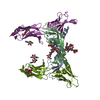


 PDBj
PDBj


 Assembly

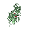
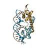
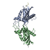
 SPODOPTERA FRUGIPERDA (fall armyworm) / Strain (production host): SF21 / References: UniProt: O43915
 Type: D-saccharide, alpha linking / Mass: 180.156 Da / Num. of mol.: 2
 Mass: 18.015 Da / Num. of mol.: 13 / Source method: isolated from a natural source / Formula: H2O
 Sample preparation
 / Beamline: X06SA / Wavelength: 1
  Processing