 Movie
Movie Controller

[English] 日本語
 Yorodumi
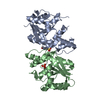
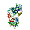
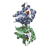
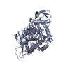
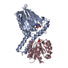
- PDB-3nb7: Crystal structure of Aquifex Aeolicus Peptidoglycan Glycosyltrans... -
+ Open data

- Basic information
| Entry | Database: PDB / ID: 3nb7 | ||||||
|---|---|---|---|---|---|---|---|
| Title | Crystal structure of Aquifex Aeolicus Peptidoglycan Glycosyltransferase in complex with Decarboxylated Neryl Moenomycin | ||||||
 Components Components | Penicillin-binding protein 1A | ||||||
 Keywords Keywords | TRANSFERASE / Glycosyltransferases / Peptidoglycan Glycosyltransferase / Polysaccharides / cell wall / antibiotics / moenomycin | ||||||
| Function / homology |  Function and homology information Function and homology informationpeptidoglycan glycosyltransferase / peptidoglycan glycosyltransferase activity / serine-type D-Ala-D-Ala carboxypeptidase / serine-type D-Ala-D-Ala carboxypeptidase activity / penicillin binding / peptidoglycan biosynthetic process / cell wall organization / regulation of cell shape / outer membrane-bounded periplasmic space / response to antibiotic ...peptidoglycan glycosyltransferase / peptidoglycan glycosyltransferase activity / serine-type D-Ala-D-Ala carboxypeptidase / serine-type D-Ala-D-Ala carboxypeptidase activity / penicillin binding / peptidoglycan biosynthetic process / cell wall organization / regulation of cell shape / outer membrane-bounded periplasmic space / response to antibiotic / proteolysis / identical protein binding / plasma membrane Similarity search - Function | ||||||
| Biological species |   Aquifex aeolicus (bacteria) Aquifex aeolicus (bacteria) | ||||||
| Method |  X-RAY DIFFRACTION / SYNCHROTRON / MOLECULAR REPLACEMENT / Resolution: 2.65 Å X-RAY DIFFRACTION / SYNCHROTRON / MOLECULAR REPLACEMENT / Resolution: 2.65 Å | ||||||
 Authors Authors | Sliz, P. / Yuan, Y. / Walker, S. | ||||||
 Citation Citation | Journal: Acs Chem.Biol. / Year: 2010 Title: Functional and structural analysis of a key region of the cell wall inhibitor moenomycin. Authors: Fuse, S. / Tsukamoto, H. / Yuan, Y. / Wang, T.S. / Zhang, Y. / Bolla, M. / Walker, S. / Sliz, P. / Kahne, D. #1: Journal: ACS CHEM.BIOL. / Year: 2008Title: Structural analysis of the contacts anchoring moenomycin to peptidoglycan glycosyltransferases and implications for antibiotic design Authors: Yuan, Y. / Fuse, S. / Ostash, B. / Sliz, P. / Kahne, D. / Walker, S. | ||||||
| History |
|
- Structure visualization
| Structure viewer | Molecule: MolmilJmol/JSmol |
|---|
- Downloads & links
-Download
| PDBx/mmCIF format | 3nb7.cif.gz | 48.1 KB | Display | PDBx/mmCIF format |
|---|---|---|---|---|
| PDB format | pdb3nb7.ent.gz | 34 KB | Display | PDB format |
| PDBx/mmJSON format | 3nb7.json.gz | Tree view | PDBx/mmJSON format | |
| Others |  Other downloads Other downloads |
-Validation report
| Arichive directory | https://data.pdbj.org/pub/pdb/validation_reports/nb/3nb7ftp://data.pdbj.org/pub/pdb/validation_reports/nb/3nb7 | HTTPS FTP |
|---|
-Related structure data
| Related structure data |  3nb6C  2oqoS S: Starting model for refinement C: citing same article ( |
|---|---|
| Similar structure data |


-Links
 PDBj
PDBj
- Assembly
| Deposited unit | 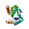
| ||||||||
|---|---|---|---|---|---|---|---|---|---|
| 1 | 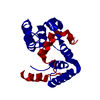
| ||||||||
| Unit cell |
|
-Components
| #1: Protein | Mass: 22912.428 Da / Num. of mol.: 1 / Fragment: UNP residues 59-243 Source method: isolated from a genetically manipulated source Source: (gene. exp.) Aquifex aeolicus (bacteria) / Strain: vf5 / Gene: aq_624, mrcA, ponA / Plasmid: pET48 b(+) / Production host: References: UniProt: O66874, Transferases; Glycosyltransferases; Pentosyltransferases |
|---|---|
| Nonpolymer details | METHYLPHOS |
-Experimental details
-Experiment
| Experiment | Method: X-RAY DIFFRACTION / Number of used crystals: 1 |
|---|
- Sample preparation
| Crystal | Density Matthews: 3.28 Å3/Da / Density % sol: 62.54 % |
|---|---|
| Crystal grow | Temperature: 295 K / Method: vapor diffusion, hanging drop / pH: 7.5 Details: 100 MM HEPES, 6% PEG6K, PH 7.5, VAPOR DIFFUSION, HANGING DROP, temperature 295K |
-Data collection
| Diffraction source | Source: SYNCHROTRON / Site: APS  / Beamline: 24-ID-C / Beamline: 24-ID-C |
|---|---|
| Detector | Detector: CCD |
| Radiation | Protocol: SINGLE WAVELENGTH / Monochromatic (M) / Laue (L): M / Scattering type: x-ray |
| Radiation wavelength | Relative weight: 1 |
| Reflection | Resolution: 2.65→50 Å / Num. obs: 8408 / % possible obs: 99.2 % / Biso Wilson estimate: 17.4 Å2 / Rsym value: 0.061 |
- Processing
| Software |
| ||||||||||||||||||||||||||||||||||||||||||||||||||||||||||||||||||||||||||||||||
|---|---|---|---|---|---|---|---|---|---|---|---|---|---|---|---|---|---|---|---|---|---|---|---|---|---|---|---|---|---|---|---|---|---|---|---|---|---|---|---|---|---|---|---|---|---|---|---|---|---|---|---|---|---|---|---|---|---|---|---|---|---|---|---|---|---|---|---|---|---|---|---|---|---|---|---|---|---|---|---|---|---|
| Refinement | Method to determine structure: MOLECULAR REPLACEMENT Starting model: PDB entry 2OQO Resolution: 2.65→25.38 Å / Rfactor Rfree error: 0.012 / Data cutoff high absF: 125502.46 / Data cutoff low absF: 0 / Isotropic thermal model: RESTRAINED / Cross valid method: THROUGHOUT / σ(F): 0 Details: BULK SOLVENT MODEL USED. THE STRUCTURE OF DECARBOXYLATED NERYL MOENOMYCIN IS NOT MODELED IN BECAUSE OF LOW OCCUPANCY, BUT THE ELECTRON DENSITY MAP CLEARLY SHOWS THE DENSITY OF THE LIGAND, ...Details: BULK SOLVENT MODEL USED. THE STRUCTURE OF DECARBOXYLATED NERYL MOENOMYCIN IS NOT MODELED IN BECAUSE OF LOW OCCUPANCY, BUT THE ELECTRON DENSITY MAP CLEARLY SHOWS THE DENSITY OF THE LIGAND, ESPECIALLY THE PHOSPHATE ATOM, IN THE LIGAND BINDING SITE
| ||||||||||||||||||||||||||||||||||||||||||||||||||||||||||||||||||||||||||||||||
| Solvent computation | Solvent model: FLAT MODEL / Bsol: 46.3539 Å2 / ksol: 0.35 e/Å3 | ||||||||||||||||||||||||||||||||||||||||||||||||||||||||||||||||||||||||||||||||
| Displacement parameters | Biso mean: 74.1 Å2
| ||||||||||||||||||||||||||||||||||||||||||||||||||||||||||||||||||||||||||||||||
| Refine analyze |
| ||||||||||||||||||||||||||||||||||||||||||||||||||||||||||||||||||||||||||||||||
| Refinement step | Cycle: LAST / Resolution: 2.65→25.38 Å
| ||||||||||||||||||||||||||||||||||||||||||||||||||||||||||||||||||||||||||||||||
| Refine LS restraints |
| ||||||||||||||||||||||||||||||||||||||||||||||||||||||||||||||||||||||||||||||||
| Refine LS restraints NCS | NCS model details: NONE | ||||||||||||||||||||||||||||||||||||||||||||||||||||||||||||||||||||||||||||||||
| LS refinement shell | Resolution: 2.65→2.82 Å / Rfactor Rfree error: 0.045 / Total num. of bins used: 6
| ||||||||||||||||||||||||||||||||||||||||||||||||||||||||||||||||||||||||||||||||
| Xplor file |
|
