 Movie
Movie Controller

[English] 日本語
 Yorodumi
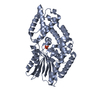
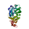
- PDB-2ps1: S. cerevisiae orotate phosphoribosyltransferase complexed with or... -
+ Open data

- Basic information
| Entry | Database: PDB / ID: 2ps1 | ||||||
|---|---|---|---|---|---|---|---|
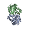
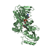
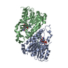
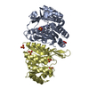
| Title | S. cerevisiae orotate phosphoribosyltransferase complexed with orotic acid and PRPP | ||||||
 Components Components | Orotate phosphoribosyltransferase 1 | ||||||
 Keywords Keywords | TRANSFERASE / ALPHA BETA / OPRTase-OA-PRPP complex | ||||||
| Function / homology |  Function and homology information Function and homology informationpyrimidine ribonucleoside biosynthetic process / orotate phosphoribosyltransferase / orotate phosphoribosyltransferase activity / pyrimidine nucleotide biosynthetic process / 'de novo' UMP biosynthetic process / 'de novo' pyrimidine nucleobase biosynthetic process / nucleus / cytoplasm Similarity search - Function | ||||||
| Biological species |  | ||||||
| Method |  X-RAY DIFFRACTION / SYNCHROTRON / MOLECULAR REPLACEMENT / Resolution: 1.75 Å X-RAY DIFFRACTION / SYNCHROTRON / MOLECULAR REPLACEMENT / Resolution: 1.75 Å | ||||||
 Authors Authors | Gonzalez-Segura, L. / Hurley, T.D. / McClard, R.W. | ||||||
 Citation Citation | Journal: Biochemistry / Year: 2007 Title: Ternary complex formation and induced asymmetry in orotate phosphoribosyltransferase. Authors: Gonzalez-Segura, L. / Witte, J.F. / McClard, R.W. / Hurley, T.D. | ||||||
| History |
|
- Structure visualization
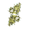
| Structure viewer | Molecule: MolmilJmol/JSmol |
|---|
- Downloads & links
-Download
| PDBx/mmCIF format | 2ps1.cif.gz | 107 KB | Display | PDBx/mmCIF format |
|---|---|---|---|---|
| PDB format | pdb2ps1.ent.gz | 80.7 KB | Display | PDB format |
| PDBx/mmJSON format | 2ps1.json.gz | Tree view | PDBx/mmJSON format | |
| Others |  Other downloads Other downloads |
-Validation report
| Arichive directory | https://data.pdbj.org/pub/pdb/validation_reports/ps/2ps1ftp://data.pdbj.org/pub/pdb/validation_reports/ps/2ps1 | HTTPS FTP |
|---|
-Related structure data
| Related structure data |  2pryC 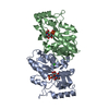 2przSC C: citing same article ( S: Starting model for refinement |
|---|---|
| Similar structure data |

-Links
 PDBj
PDBj
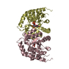
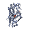
- Assembly
| Deposited unit | 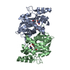
| ||||||||
|---|---|---|---|---|---|---|---|---|---|
| 1 |
| ||||||||
| Unit cell |
|
-Components
| #1: Protein | Mass: 24689.439 Da / Num. of mol.: 2 Source method: isolated from a genetically manipulated source Source: (gene. exp.) Gene: URA5, PYR5 / Plasmid: pREJ2 / Production host:  References: UniProt: P13298, orotate phosphoribosyltransferase #2: Chemical |   Mass: 24.305 Da / Num. of mol.: 2 / Source method: obtained synthetically / Formula: Mg Mass: 24.305 Da / Num. of mol.: 2 / Source method: obtained synthetically / Formula: Mg#3: Chemical | 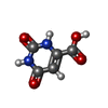  Mass: 156.096 Da / Num. of mol.: 2 / Source method: obtained synthetically / Formula: C5H4N2O4 Mass: 156.096 Da / Num. of mol.: 2 / Source method: obtained synthetically / Formula: C5H4N2O4#4: Sugar |   Type: D-saccharide / Mass: 390.070 Da / Num. of mol.: 2 / Source method: obtained synthetically / Formula: C5H13O14P3 Type: D-saccharide / Mass: 390.070 Da / Num. of mol.: 2 / Source method: obtained synthetically / Formula: C5H13O14P3#5: Water | ChemComp-HOH / |  Mass: 18.015 Da / Num. of mol.: 322 / Source method: isolated from a natural source / Formula: H2O Mass: 18.015 Da / Num. of mol.: 322 / Source method: isolated from a natural source / Formula: H2O |
|---|
-Experimental details
-Experiment
| Experiment | Method: X-RAY DIFFRACTION / Number of used crystals: 1 |
|---|
- Sample preparation
| Crystal | Density Matthews: 2.04 Å3/Da / Density % sol: 39.6 % |
|---|---|
| Crystal grow | Temperature: 288.15 K / Method: vapor diffusion, sitting drop / pH: 4.6 Details: 38% PEG 4000, 0.14M ammonium acetate, 0.1M sodium acetate, 2.0mM magnesium chloride, 5.0mM PRPP, 5.0mM orotic acid, pH 4.6, VAPOR DIFFUSION, SITTING DROP, temperature 288.15K |
-Data collection
| Diffraction | Mean temperature: 100 K |
|---|---|
| Diffraction source | Source: SYNCHROTRON / Site: APS  / Beamline: 19-ID / Wavelength: 0.98 Å / Beamline: 19-ID / Wavelength: 0.98 Å |
| Detector | Type: ADSC QUANTUM 315 / Detector: CCD / Date: Apr 1, 2006 |
| Radiation | Protocol: SINGLE WAVELENGTH / Monochromatic (M) / Laue (L): M / Scattering type: x-ray |
| Radiation wavelength | Wavelength: 0.98 Å / Relative weight: 1 |
| Reflection | Resolution: 1.75→50 Å / Num. all: 39461 / Num. obs: 36660 / % possible obs: 92.9 % / Observed criterion σ(F): 0 / Observed criterion σ(I): 0.2 / Redundancy: 3.6 % / Biso Wilson estimate: 21.245 Å2 / Rmerge(I) obs: 0.049 / Rsym value: 0.043 / Χ2: 1.064 / Net I/σ(I): 17.8 |
| Reflection shell | Resolution: 1.75→1.81 Å / Redundancy: 2.5 % / Rmerge(I) obs: 0.251 / Mean I/σ(I) obs: 3.2 / Num. unique all: 2533 / Rsym value: 0.211 / Χ2: 0.944 / % possible all: 63.7 |
- Processing
| Software |
| ||||||||||||||||||||||||||||||||||||||||||||||||||||||||||||||||||||||||||||||||||||||||||
|---|---|---|---|---|---|---|---|---|---|---|---|---|---|---|---|---|---|---|---|---|---|---|---|---|---|---|---|---|---|---|---|---|---|---|---|---|---|---|---|---|---|---|---|---|---|---|---|---|---|---|---|---|---|---|---|---|---|---|---|---|---|---|---|---|---|---|---|---|---|---|---|---|---|---|---|---|---|---|---|---|---|---|---|---|---|---|---|---|---|---|---|
| Refinement | Method to determine structure: MOLECULAR REPLACEMENT Starting model: PDB ENTRY 2PRZ (S. cerevisiae OPRTase complexed with OMP) Resolution: 1.75→49.94 Å / Cor.coef. Fo:Fc: 0.954 / Cor.coef. Fo:Fc free: 0.946 / SU B: 2.715 / SU ML: 0.089 / Cross valid method: THROUGHOUT / σ(F): 0 / σ(I): 0 / ESU R: 0.157 / ESU R Free: 0.131 / Stereochemistry target values: MAXIMUM LIKELIHOOD / Details: HYDROGENS HAVE BEEN ADDED IN THE RIDING POSITIONS
| ||||||||||||||||||||||||||||||||||||||||||||||||||||||||||||||||||||||||||||||||||||||||||
| Solvent computation | Ion probe radii: 0.8 Å / Shrinkage radii: 0.8 Å / VDW probe radii: 1.2 Å / Solvent model: MASK | ||||||||||||||||||||||||||||||||||||||||||||||||||||||||||||||||||||||||||||||||||||||||||
| Displacement parameters | Biso mean: 23.628 Å2
| ||||||||||||||||||||||||||||||||||||||||||||||||||||||||||||||||||||||||||||||||||||||||||
| Refinement step | Cycle: LAST / Resolution: 1.75→49.94 Å
| ||||||||||||||||||||||||||||||||||||||||||||||||||||||||||||||||||||||||||||||||||||||||||
| Refine LS restraints |
| ||||||||||||||||||||||||||||||||||||||||||||||||||||||||||||||||||||||||||||||||||||||||||
| LS refinement shell | Resolution: 1.75→1.792 Å / Total num. of bins used: 20
|
