 Movie
Movie Controller

[English] 日本語
 Yorodumi
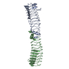
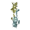
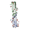
- PDB-2xtw: Structure of QnrB1 (Full length), a plasmid-mediated fluoroquinol... -
+ Open data

- Basic information
| Entry | Database: PDB / ID: 2xtw | ||||||
|---|---|---|---|---|---|---|---|
| Title | Structure of QnrB1 (Full length), a plasmid-mediated fluoroquinolone resistance protein | ||||||
 Components Components | QNRB1 | ||||||
 Keywords Keywords | CELL CYCLE / PENTAPEPTIDE REPEAT / PRP / ANTIBIOTIC RESISTANCE / RIGHT HANDED QUADRILATERAL BETA-HELIX | ||||||
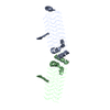
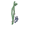
| Function / homology | Pentapeptide repeat region / : / Pentapeptide repeats (8 copies) / Pentapeptide repeat / E3 ubiquitin-protein ligase SopA / Pectate Lyase C-like / 3 Solenoid / Mainly Beta / QnrB1 Function and homology information Function and homology information | ||||||
| Biological species |  KLEBSIELLA PNEUMONIAE (bacteria) KLEBSIELLA PNEUMONIAE (bacteria) | ||||||
| Method |  X-RAY DIFFRACTION / MOLECULAR REPLACEMENT / Resolution: 2.803 Å X-RAY DIFFRACTION / MOLECULAR REPLACEMENT / Resolution: 2.803 Å | ||||||
 Authors Authors | Vetting, M.W. / Hegde, S.S. / Park, C.H. / Jacoby, G.A. / Hooper, D.C. / Blanchard, J.S. | ||||||
 Citation Citation | Journal: J.Biol.Chem. / Year: 2011 Title: Structure of Qnrb1, a Plasmid-Mediated Fluoroquinolone Resistance Factor. Authors: Vetting, M.W. / Hegde, S.S. / Wang, M. / Jacoby, G.A. / Hooper, D.C. / Blanchard, J.S. | ||||||
| History |
|
- Structure visualization
| Structure viewer | Molecule: MolmilJmol/JSmol |
|---|
- Downloads & links
-Download
| PDBx/mmCIF format | 2xtw.cif.gz | 166.5 KB | Display | PDBx/mmCIF format |
|---|---|---|---|---|
| PDB format | pdb2xtw.ent.gz | 134.9 KB | Display | PDB format |
| PDBx/mmJSON format | 2xtw.json.gz | Tree view | PDBx/mmJSON format | |
| Others |  Other downloads Other downloads |
-Validation report
| Summary document | 2xtw_validation.pdf.gz | 445.4 KB | Display | wwPDB validaton report |
|---|---|---|---|---|
| Full document | 2xtw_full_validation.pdf.gz | 461.9 KB | Display | |
| Data in XML | 2xtw_validation.xml.gz | 30.5 KB | Display | |
| Data in CIF | 2xtw_validation.cif.gz | 41 KB | Display | |
| Arichive directory | https://data.pdbj.org/pub/pdb/validation_reports/xt/2xtwftp://data.pdbj.org/pub/pdb/validation_reports/xt/2xtw | HTTPS FTP |
-Related structure data


-Links
 PDBj
PDBj- Assembly
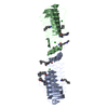
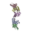
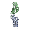
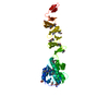
| Deposited unit | 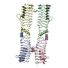
| ||||||||
|---|---|---|---|---|---|---|---|---|---|
| 1 | 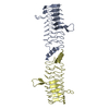
| ||||||||
| 2 | 
| ||||||||
| Unit cell |
|
-Components
| #1: Protein | Mass: 24126.078 Da / Num. of mol.: 4 Fragment: TOPISOMERASE POISON RESISTANCE FACTOR, RESIDUES 13-226 Source method: isolated from a genetically manipulated source Source: (gene. exp.) KLEBSIELLA PNEUMONIAE (bacteria) / Production host: Sequence details | CLONED INTO A CLEAVABLE PET28 VECTOR WITH CLEAVABLE HIS6 TAG. PROTEIN IS CLEAVED LEAVING GSH ON N ...CLONED INTO A CLEAVABLE PET28 VECTOR WITH CLEAVABLE HIS6 TAG. PROTEIN IS CLEAVED LEAVING GSH ON N TERMINUS. PROTEIN WAS CLONED STARTING FROM 2ND MET OF THE GENBANK SEQUENCE. | |
|---|
-Experimental details
-Experiment
| Experiment | Method: X-RAY DIFFRACTION / Number of used crystals: 1 |
|---|
- Sample preparation
| Crystal | Density Matthews: 2.73 Å3/Da / Density % sol: 48 % / Description: NONE |
|---|---|
| Crystal grow | Temperature: 277 K / pH: 4.5 Details: 100 MM NA/K PHOSPHATE PH 4.5, 2 M NACL AT 4 DEGREES C |
-Data collection
| Diffraction | Mean temperature: 93 K |
|---|---|
| Diffraction source | Source: ROTATING ANODE / Type: RIGAKU RU300 / Wavelength: 1.5418 |
| Detector | Type: RIGAKU RAXIS IV / Detector: IMAGE PLATE / Details: MIRRORS |
| Radiation | Monochromator: NI FILTER / Protocol: SINGLE WAVELENGTH / Monochromatic (M) / Laue (L): M / Scattering type: x-ray |
| Radiation wavelength | Wavelength: 1.5418 Å / Relative weight: 1 |
| Reflection | Resolution: 2.8→40 Å / Num. obs: 26120 / % possible obs: 99 % / Observed criterion σ(I): 0 / Redundancy: 7.8 % / Biso Wilson estimate: 39.58 Å2 / Rmerge(I) obs: 0.08 / Net I/σ(I): 18.7 |
| Reflection shell | Resolution: 2.8→2.9 Å / Redundancy: 7.4 % / Rmerge(I) obs: 0.28 / Mean I/σ(I) obs: 6.5 / % possible all: 98.2 |
- Processing
| Software |
| ||||||||||||||||||||||||||||||||||||||||||||||||||||||||||||||||||||||
|---|---|---|---|---|---|---|---|---|---|---|---|---|---|---|---|---|---|---|---|---|---|---|---|---|---|---|---|---|---|---|---|---|---|---|---|---|---|---|---|---|---|---|---|---|---|---|---|---|---|---|---|---|---|---|---|---|---|---|---|---|---|---|---|---|---|---|---|---|---|---|---|
| Refinement | Method to determine structure: MOLECULAR REPLACEMENT / Resolution: 2.803→47.268 Å / SU ML: 0.43 / σ(F): 0 / Phase error: 35.02 / Stereochemistry target values: ML
| ||||||||||||||||||||||||||||||||||||||||||||||||||||||||||||||||||||||
| Solvent computation | Shrinkage radii: 0.83 Å / VDW probe radii: 1.1 Å / Solvent model: FLAT BULK SOLVENT MODEL / Bsol: 21.22 Å2 / ksol: 0.366 e/Å3 | ||||||||||||||||||||||||||||||||||||||||||||||||||||||||||||||||||||||
| Displacement parameters | Biso mean: 38.1 Å2
| ||||||||||||||||||||||||||||||||||||||||||||||||||||||||||||||||||||||
| Refinement step | Cycle: LAST / Resolution: 2.803→47.268 Å
| ||||||||||||||||||||||||||||||||||||||||||||||||||||||||||||||||||||||
| Refine LS restraints |
| ||||||||||||||||||||||||||||||||||||||||||||||||||||||||||||||||||||||
| LS refinement shell |
|
