 Movie
Movie Controller

[English] 日本語
 Yorodumi
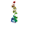
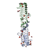
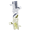
- PDB-2w7z: Structure of the pentapeptide repeat protein EfsQnr, a DNA gyrase... -
+ Open data

- Basic information
| Entry | Database: PDB / ID: 2w7z | ||||||
|---|---|---|---|---|---|---|---|
| Title | Structure of the pentapeptide repeat protein EfsQnr, a DNA gyrase inhibitor. Free amines modified by cyclic pentylation with glutaraldehyde. | ||||||
 Components Components | PENTAPEPTIDE REPEAT FAMILY PROTEIN | ||||||
 Keywords Keywords | INHIBITOR / GLUTARALDEHYDE / GYRASE INHIBITOR / CYCLIC PENTYLATION / CHEMICAL MODIFICATION / PENTAPEPTIDE REPEAT PROTEIN | ||||||
| Function / homology | : / Pentapeptide repeat region / Pentapeptide repeats (8 copies) / Pentapeptide repeat / E3 ubiquitin-protein ligase SopA / Pectate Lyase C-like / 3 Solenoid / Mainly Beta / Pentapeptide repeat family protein Function and homology information Function and homology information | ||||||
| Biological species |   ENTEROCOCCUS FAECALIS (bacteria) ENTEROCOCCUS FAECALIS (bacteria) | ||||||
| Method |  X-RAY DIFFRACTION / SAD / Resolution: 1.6 Å X-RAY DIFFRACTION / SAD / Resolution: 1.6 Å | ||||||
 Authors Authors | Vetting, M.W. / Hegde, S.S. / Blanchard, J.S. | ||||||
 Citation Citation | Journal: Acta Crystallogr.,Sect.D / Year: 2009 Title: Crystallization of a Pentapeptide-Repeat Protein by Reductive Cyclic Pentylation of Free Amines with Glutaraldehyde. Authors: Vetting, M.W. / Hegde, S.S. / Blanchard, J.S. | ||||||
| History |
| ||||||
| Remark 700 | SHEET THE SHEET STRUCTURE OF THIS MOLECULE IS BIFURCATED. IN ORDER TO REPRESENT THIS FEATURE IN ... SHEET THE SHEET STRUCTURE OF THIS MOLECULE IS BIFURCATED. IN ORDER TO REPRESENT THIS FEATURE IN THE SHEET RECORDS BELOW, TWO SHEETS ARE DEFINED. |
- Structure visualization
| Structure viewer | Molecule: MolmilJmol/JSmol |
|---|
- Downloads & links
-Download
| PDBx/mmCIF format | 2w7z.cif.gz | 107.5 KB | Display | PDBx/mmCIF format |
|---|---|---|---|---|
| PDB format | pdb2w7z.ent.gz | 84.2 KB | Display | PDB format |
| PDBx/mmJSON format | 2w7z.json.gz | Tree view | PDBx/mmJSON format | |
| Others |  Other downloads Other downloads |
-Validation report
| Arichive directory | https://data.pdbj.org/pub/pdb/validation_reports/w7/2w7zftp://data.pdbj.org/pub/pdb/validation_reports/w7/2w7z | HTTPS FTP |
|---|
-Related structure data
| Similar structure data |
|---|


-Links
 PDBj
PDBj- Assembly
| Deposited unit | 
| ||||||||
|---|---|---|---|---|---|---|---|---|---|
| 1 |
| ||||||||
| Unit cell |
| ||||||||
| Noncrystallographic symmetry (NCS) | NCS oper: (Code: given Matrix: (-0.8509, -0.5237, -0.04045), Vector: |
-Components
| #1: Protein | Mass: 25023.357 Da / Num. of mol.: 2 Source method: isolated from a genetically manipulated source Details: CHEMICAL MODIFICATION OF THE FREE AMINES (LYSINES, N-TERMINUS) BY TREATMENT WITH GLUTARALDEHYDE UNDER REDUCING CONDITIONS Source: (gene. exp.) ENTEROCOCCUS FAECALIS (bacteria) / Plasmid: PET28 / Production host: #2: Chemical | ChemComp-CL /   Mass: 35.453 Da / Num. of mol.: 4 / Source method: obtained synthetically / Formula: Cl Mass: 35.453 Da / Num. of mol.: 4 / Source method: obtained synthetically / Formula: Cl#3: Water | ChemComp-HOH / |  Mass: 18.015 Da / Num. of mol.: 417 / Source method: isolated from a natural source / Formula: H2O Mass: 18.015 Da / Num. of mol.: 417 / Source method: isolated from a natural source / Formula: H2OHas protein modification | Y | Nonpolymer details | LYSYL-PIPERIDINE (LPD): UNSATURATED SIX-MEMBERED RING, WITH PROTONATED NITROGEN, SP3 GEOMETRY, AND ...LYSYL-PIPERIDINE | |
|---|
-Experimental details
-Experiment
| Experiment | Method: X-RAY DIFFRACTION / Number of used crystals: 1 |
|---|
- Sample preparation
| Crystal | Density Matthews: 2 Å3/Da / Density % sol: 38.14 % / Description: NONE |
|---|---|
| Crystal grow | pH: 6 Details: PROTEIN - 10 MG/ML, 400 MM AMMONIUM SULFATE, 15 MM HEPES PH 7.5, 1 MM DTT, 1 MM ETDA PRECIPITANT - 100 MM MES PH 6.5, 5-15% PEG 6000, 1M LICL |
-Data collection
| Diffraction | Mean temperature: 100 K |
|---|---|
| Diffraction source | Source: ROTATING ANODE / Type: RIGAKU RUH3R / Wavelength: 1.5418 |
| Detector | Type: RIGAKU R-AXIS IV / Date: Jan 2, 2008 |
| Radiation | Protocol: SINGLE WAVELENGTH / Monochromatic (M) / Laue (L): M / Scattering type: x-ray |
| Radiation wavelength | Wavelength: 1.5418 Å / Relative weight: 1 |
| Reflection | Resolution: 1.6→20.6 Å / Num. obs: 53909 / % possible obs: 94.7 % / Observed criterion σ(I): 0 / Redundancy: 2.9 % / Rmerge(I) obs: 0.04 / Net I/σ(I): 19.3 |
| Reflection shell | Resolution: 1.6→1.69 Å / Redundancy: 2.7 % / Rmerge(I) obs: 0.25 / Mean I/σ(I) obs: 3.1 / % possible all: 86 |
- Processing
| Software |
| ||||||||||||||||||||||||||||||||||||||||||||||||||||||||||||||||||||||||||||||||||||||||||||||||||||||||||||||||||||||||||||||||||||||||||||||||||||||||||||||||||||||||||||||||||||||
|---|---|---|---|---|---|---|---|---|---|---|---|---|---|---|---|---|---|---|---|---|---|---|---|---|---|---|---|---|---|---|---|---|---|---|---|---|---|---|---|---|---|---|---|---|---|---|---|---|---|---|---|---|---|---|---|---|---|---|---|---|---|---|---|---|---|---|---|---|---|---|---|---|---|---|---|---|---|---|---|---|---|---|---|---|---|---|---|---|---|---|---|---|---|---|---|---|---|---|---|---|---|---|---|---|---|---|---|---|---|---|---|---|---|---|---|---|---|---|---|---|---|---|---|---|---|---|---|---|---|---|---|---|---|---|---|---|---|---|---|---|---|---|---|---|---|---|---|---|---|---|---|---|---|---|---|---|---|---|---|---|---|---|---|---|---|---|---|---|---|---|---|---|---|---|---|---|---|---|---|---|---|---|---|
| Refinement | Method to determine structure: SAD Starting model: NONE Resolution: 1.6→94.07 Å / Cor.coef. Fo:Fc: 0.96 / Cor.coef. Fo:Fc free: 0.942 / SU B: 3.462 / SU ML: 0.063 / TLS residual ADP flag: LIKELY RESIDUAL / Cross valid method: THROUGHOUT / ESU R: 0.105 / ESU R Free: 0.102 / Stereochemistry target values: MAXIMUM LIKELIHOOD Details: HYDROGENS HAVE BEEN ADDED IN THE RIDING POSITIONS. PROTEIN WAS PRODUCED WITH N-TERMINAL THROMBIN CLEAVABLE TAG. PROTEIN WAS CRYSTALLIZED AFTER THROMBIN CLEAVAGE, LEAVING A NON-NATIVE N-TERMINUS OF GLYSERHIS-MET1
| ||||||||||||||||||||||||||||||||||||||||||||||||||||||||||||||||||||||||||||||||||||||||||||||||||||||||||||||||||||||||||||||||||||||||||||||||||||||||||||||||||||||||||||||||||||||
| Solvent computation | Ion probe radii: 0.8 Å / Shrinkage radii: 0.8 Å / VDW probe radii: 1.2 Å / Solvent model: MASK | ||||||||||||||||||||||||||||||||||||||||||||||||||||||||||||||||||||||||||||||||||||||||||||||||||||||||||||||||||||||||||||||||||||||||||||||||||||||||||||||||||||||||||||||||||||||
| Displacement parameters | Biso mean: 15.48 Å2
| ||||||||||||||||||||||||||||||||||||||||||||||||||||||||||||||||||||||||||||||||||||||||||||||||||||||||||||||||||||||||||||||||||||||||||||||||||||||||||||||||||||||||||||||||||||||
| Refinement step | Cycle: LAST / Resolution: 1.6→94.07 Å
| ||||||||||||||||||||||||||||||||||||||||||||||||||||||||||||||||||||||||||||||||||||||||||||||||||||||||||||||||||||||||||||||||||||||||||||||||||||||||||||||||||||||||||||||||||||||
| Refine LS restraints |
|