 Movie
Movie Controller

[English] 日本語
 Yorodumi
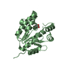
- PDB-2xpo: Crystal structure of a Spt6-Iws1(Spn1) complex from Encephalitozo... -
+ Open data

- Basic information
| Entry | Database: PDB / ID: 2xpo | ||||||
|---|---|---|---|---|---|---|---|
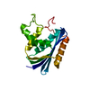
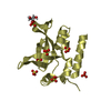
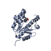
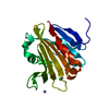
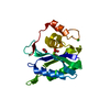
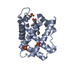
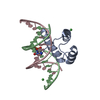
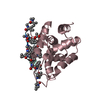
| Title | Crystal structure of a Spt6-Iws1(Spn1) complex from Encephalitozoon cuniculi, Form II | ||||||
 Components Components |
| ||||||
 Keywords Keywords | TRANSCRIPTION / ELONGATION / HISTONE CHAPERONE / RNA POLYMERASE II / MRNA EXPORT | ||||||
| Function / homology |  Function and homology information Function and homology informationnucleosome organization / transcription elongation-coupled chromatin remodeling / poly(A)+ mRNA export from nucleus / nucleosome binding / transcription elongation factor complex / histone binding / nucleus Similarity search - Function | ||||||
| Biological species |  ENCEPHALITOZOON CUNICULI (fungus) ENCEPHALITOZOON CUNICULI (fungus) | ||||||
| Method |  X-RAY DIFFRACTION / SYNCHROTRON / MOLECULAR REPLACEMENT / Resolution: 2.1 Å X-RAY DIFFRACTION / SYNCHROTRON / MOLECULAR REPLACEMENT / Resolution: 2.1 Å | ||||||
 Authors Authors | Diebold, M.-L. / Koch, M. / Cura, V. / Moras, D. / Cavarelli, J. / Romier, C. | ||||||
 Citation Citation | Journal: Embo J. / Year: 2010 Title: The Structure of an Iws1/Spt6 Complex Reveals an Interaction Domain Conserved in Tfiis, Elongin a and Med26 Authors: Diebold, M.-L. / Koch, M. / Loeliger, E. / Cura, V. / Winston, F. / Cavarelli, J. / Romier, C. | ||||||
| History |
|
- Structure visualization
| Structure viewer | Molecule: MolmilJmol/JSmol |
|---|
- Downloads & links
-Download
| PDBx/mmCIF format | 2xpo.cif.gz | 141 KB | Display | PDBx/mmCIF format |
|---|---|---|---|---|
| PDB format | pdb2xpo.ent.gz | 112 KB | Display | PDB format |
| PDBx/mmJSON format | 2xpo.json.gz | Tree view | PDBx/mmJSON format | |
| Others |  Other downloads Other downloads |
-Validation report
| Arichive directory | https://data.pdbj.org/pub/pdb/validation_reports/xp/2xpoftp://data.pdbj.org/pub/pdb/validation_reports/xp/2xpo | HTTPS FTP |
|---|
-Related structure data
| Related structure data | 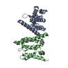 2xplSC 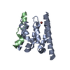 2xpnC 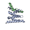 2xppC C: citing same article ( S: Starting model for refinement |
|---|---|
| Similar structure data |

-Links
 PDBj
PDBj- Assembly
| Deposited unit | 
| ||||||||
|---|---|---|---|---|---|---|---|---|---|
| 1 | 
| ||||||||
| 2 | 
| ||||||||
| Unit cell |
|
-Components
| #1: Protein | Mass: 16424.105 Da / Num. of mol.: 2 / Fragment: EVOLUTIONARY CONSERVED DOMAIN, RESIDUES 55-198 Source method: isolated from a genetically manipulated source Source: (gene. exp.) ENCEPHALITOZOON CUNICULI (fungus) / Plasmid: PNCS / Production host:  #2: Protein/peptide | Mass: 2712.939 Da / Num. of mol.: 2 / Fragment: N-TERMINAL FRAGMENT, RESIDUES 53-71 Source method: isolated from a genetically manipulated source Source: (gene. exp.) ENCEPHALITOZOON CUNICULI (fungus) / Plasmid: PNEA-TH / Production host: #3: Chemical |   Mass: 35.453 Da / Num. of mol.: 2 / Source method: obtained synthetically / Formula: Cl Mass: 35.453 Da / Num. of mol.: 2 / Source method: obtained synthetically / Formula: Cl#4: Water | ChemComp-HOH / |  Mass: 18.015 Da / Num. of mol.: 151 / Source method: isolated from a natural source / Formula: H2O Mass: 18.015 Da / Num. of mol.: 151 / Source method: isolated from a natural source / Formula: H2OSequence details | B, D 49-50 PART OF THROMBIN CLEAVAGE SITE. B, D 51-52 PART OF NDEI CLONING SITE. | |
|---|
-Experimental details
-Experiment
| Experiment | Method: X-RAY DIFFRACTION / Number of used crystals: 1 |
|---|
- Sample preparation
| Crystal | Density Matthews: 2.46 Å3/Da / Density % sol: 50 % / Description: NONE |
|---|---|
| Crystal grow | pH: 5 / Details: 0.1 M SODIUM ACETATE 18% PEG1500 0.05 M MGCL2 |
-Data collection
| Diffraction | Mean temperature: 100 K |
|---|---|
| Diffraction source | Source: SYNCHROTRON / Site: ESRF  / Beamline: ID14-1 / Wavelength: 0.9334 / Beamline: ID14-1 / Wavelength: 0.9334 |
| Detector | Type: ADSC CCD / Detector: CCD / Date: Dec 11, 2008 |
| Radiation | Protocol: SINGLE WAVELENGTH / Monochromatic (M) / Laue (L): M / Scattering type: x-ray |
| Radiation wavelength | Wavelength: 0.9334 Å / Relative weight: 1 |
| Reflection | Resolution: 2.1→50 Å / Num. obs: 21935 / % possible obs: 99.9 % / Observed criterion σ(I): 2 / Redundancy: 10.7 % / Rmerge(I) obs: 0.08 / Net I/σ(I): 33.6 |
| Reflection shell | Resolution: 2.1→2.15 Å / Redundancy: 10.2 % / Rmerge(I) obs: 0.29 / Mean I/σ(I) obs: 10.47 / % possible all: 100 |
- Processing
| Software |
| ||||||||||||||||||||||||||||||||||||||||||||||||||||||||||||||||||||||||||||||||||||||||||||||||||||||||||||||||||||||||||||||||||||||||||||||||||||||||||||||||||||||||||||||||||||||
|---|---|---|---|---|---|---|---|---|---|---|---|---|---|---|---|---|---|---|---|---|---|---|---|---|---|---|---|---|---|---|---|---|---|---|---|---|---|---|---|---|---|---|---|---|---|---|---|---|---|---|---|---|---|---|---|---|---|---|---|---|---|---|---|---|---|---|---|---|---|---|---|---|---|---|---|---|---|---|---|---|---|---|---|---|---|---|---|---|---|---|---|---|---|---|---|---|---|---|---|---|---|---|---|---|---|---|---|---|---|---|---|---|---|---|---|---|---|---|---|---|---|---|---|---|---|---|---|---|---|---|---|---|---|---|---|---|---|---|---|---|---|---|---|---|---|---|---|---|---|---|---|---|---|---|---|---|---|---|---|---|---|---|---|---|---|---|---|---|---|---|---|---|---|---|---|---|---|---|---|---|---|---|---|
| Refinement | Method to determine structure: MOLECULAR REPLACEMENT Starting model: PDB ENTRY 2XPL Resolution: 2.1→97.5 Å / Cor.coef. Fo:Fc: 0.936 / Cor.coef. Fo:Fc free: 0.903 / SU B: 9.188 / SU ML: 0.115 / Cross valid method: THROUGHOUT / ESU R: 0.203 / ESU R Free: 0.181 / Stereochemistry target values: MAXIMUM LIKELIHOOD Details: HYDROGENS HAVE BEEN ADDED IN THE RIDING POSITIONS. U VALUES WITH TLS ADDED. ATOM RECORD CONTAINS SUM OF TLS AND RESIDUAL B FACTORS.
| ||||||||||||||||||||||||||||||||||||||||||||||||||||||||||||||||||||||||||||||||||||||||||||||||||||||||||||||||||||||||||||||||||||||||||||||||||||||||||||||||||||||||||||||||||||||
| Solvent computation | Ion probe radii: 0.8 Å / Shrinkage radii: 0.8 Å / VDW probe radii: 1.4 Å / Solvent model: MASK | ||||||||||||||||||||||||||||||||||||||||||||||||||||||||||||||||||||||||||||||||||||||||||||||||||||||||||||||||||||||||||||||||||||||||||||||||||||||||||||||||||||||||||||||||||||||
| Displacement parameters | Biso mean: 24.14 Å2
| ||||||||||||||||||||||||||||||||||||||||||||||||||||||||||||||||||||||||||||||||||||||||||||||||||||||||||||||||||||||||||||||||||||||||||||||||||||||||||||||||||||||||||||||||||||||
| Refinement step | Cycle: LAST / Resolution: 2.1→97.5 Å
| ||||||||||||||||||||||||||||||||||||||||||||||||||||||||||||||||||||||||||||||||||||||||||||||||||||||||||||||||||||||||||||||||||||||||||||||||||||||||||||||||||||||||||||||||||||||
| Refine LS restraints |
|