 Movie
Movie Controller

[English] 日本語
 Yorodumi
- PDB-2xn5: Crystal structure of thyroxine-binding globulin complexed with Fu... -
+ Open data

- Basic information
| Entry | Database: PDB / ID: 2xn5 | ||||||
|---|---|---|---|---|---|---|---|
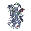
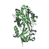
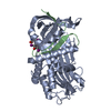
| Title | Crystal structure of thyroxine-binding globulin complexed with Furosemide | ||||||
 Components Components | (THYROXINE-BINDING ...) x 2 | ||||||
 Keywords Keywords | TRANSPORT / CLEAVED PROTEIN | ||||||
| Function / homology |  Function and homology information Function and homology informationthyroid hormone transport / serine-type endopeptidase inhibitor activity / : / extracellular exosome / extracellular region Similarity search - Function | ||||||
| Biological species |  HOMO SAPIENS (human) HOMO SAPIENS (human) | ||||||
| Method |  X-RAY DIFFRACTION / SYNCHROTRON / MOLECULAR REPLACEMENT / Resolution: 1.7 Å X-RAY DIFFRACTION / SYNCHROTRON / MOLECULAR REPLACEMENT / Resolution: 1.7 Å | ||||||
 Authors Authors | Qi, X. / Yan, Y. / Wei, Z. / Zhou, A. | ||||||
 Citation Citation | Journal: J.Biol.Chem. / Year: 2011 Title: Allosteric Modulation of Hormone Release from Thyroxine and Corticosteroid Binding-Globulins. Authors: Qi, X. / Loiseau, F. / Chan, W.L. / Yan, Y. / Wei, Z. / Milroy, L.G. / Myers, R.M. / Ley, S.V. / Read, R.J. / Carrell, R.W. / Zhou, A. | ||||||
| History |
|
- Structure visualization
| Structure viewer | Molecule: MolmilJmol/JSmol |
|---|
- Downloads & links
-Download
| PDBx/mmCIF format | 2xn5.cif.gz | 168.8 KB | Display | PDBx/mmCIF format |
|---|---|---|---|---|
| PDB format | pdb2xn5.ent.gz | 131.4 KB | Display | PDB format |
| PDBx/mmJSON format | 2xn5.json.gz | Tree view | PDBx/mmJSON format | |
| Others |  Other downloads Other downloads |
-Validation report
| Arichive directory | https://data.pdbj.org/pub/pdb/validation_reports/xn/2xn5ftp://data.pdbj.org/pub/pdb/validation_reports/xn/2xn5 | HTTPS FTP |
|---|
-Related structure data
| Related structure data |  2rivSC  2riwC  2xn3C  2xn6C 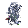 2xn7C C: citing same article ( S: Starting model for refinement |
|---|---|
| Similar structure data |


-Links
 PDBj
PDBj
- Assembly
| Deposited unit | 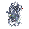
| ||||||||
|---|---|---|---|---|---|---|---|---|---|
| 1 |
| ||||||||
| Unit cell |
| ||||||||
| Components on special symmetry positions |
|
-Components
-THYROXINE-BINDING ... , 2 types, 2 molecules AB
| #1: Protein | Mass: 39145.879 Da / Num. of mol.: 1 / Fragment: RESIDUES 32-380 / Mutation: YES Source method: isolated from a genetically manipulated source Details: RESIDUES 346-355 OF TBG WERE REPLACED BY GAMFLEAIPRS Source: (gene. exp.) HOMO SAPIENS (human) / Production host:  |
|---|---|
| #2: Protein/peptide | Mass: 4026.811 Da / Num. of mol.: 1 / Fragment: RESIDUES 381-415 Source method: isolated from a genetically manipulated source Source: (gene. exp.) HOMO SAPIENS (human) / Production host: |
-Non-polymers , 4 types, 282 molecules 


| #3: Chemical | ChemComp-FUN /  Mass: 330.744 Da / Num. of mol.: 1 / Source method: obtained synthetically / Formula: C12H11ClN2O5S Mass: 330.744 Da / Num. of mol.: 1 / Source method: obtained synthetically / Formula: C12H11ClN2O5S | ||||
|---|---|---|---|---|---|
| #4: Chemical | ChemComp-CA /  Mass: 40.078 Da / Num. of mol.: 4 / Source method: obtained synthetically / Formula: Ca Mass: 40.078 Da / Num. of mol.: 4 / Source method: obtained synthetically / Formula: Ca#5: Chemical | ChemComp-EDO /  Mass: 62.068 Da / Num. of mol.: 4 / Source method: obtained synthetically / Formula: C2H6O2 Mass: 62.068 Da / Num. of mol.: 4 / Source method: obtained synthetically / Formula: C2H6O2#6: Water | ChemComp-HOH / | Mass: 18.015 Da / Num. of mol.: 273 / Source method: isolated from a natural source / Formula: H2O |
-Details
| Compound details | ENGINEERED RESIDUE IN CHAIN A, ALA 366 TO GLY ENGINEERED RESIDUE IN CHAIN A, VAL 367 TO ALA ...ENGINEERED |
|---|
-Experimental details
-Experiment
| Experiment | Method: X-RAY DIFFRACTION / Number of used crystals: 1 |
|---|
- Sample preparation
| Crystal | Density Matthews: 2.35 Å3/Da / Density % sol: 48 % / Description: NONE |
|---|---|
| Crystal grow | pH: 5.4 / Details: 20% PEG3350, pH 5.4 |
-Data collection
| Diffraction | Mean temperature: 100 K |
|---|---|
| Diffraction source | Source: SYNCHROTRON / Site: Diamond  / Beamline: I02 / Wavelength: 0.998 / Beamline: I02 / Wavelength: 0.998 |
| Radiation | Protocol: SINGLE WAVELENGTH / Monochromatic (M) / Laue (L): M / Scattering type: x-ray |
| Radiation wavelength | Wavelength: 0.998 Å / Relative weight: 1 |
| Reflection | Resolution: 1.7→34.58 Å / Num. obs: 40382 / % possible obs: 92.6 % / Observed criterion σ(I): 2 / Redundancy: 3.7 % / Rmerge(I) obs: 0.19 / Net I/σ(I): 4.3 |
| Reflection shell | Resolution: 1.7→1.79 Å / Redundancy: 3.6 % / Rmerge(I) obs: 0.55 / Mean I/σ(I) obs: 1.7 / % possible all: 71.1 |
- Processing
| Software |
| ||||||||||||||||||||||||||||||||||||||||||||||||||||||||||||||||||||||||||||||||||||||||||||||||||||||||||||||||||||||||||||||||||||||||||||||||||||||||||||||||||||||||||||||||||||||
|---|---|---|---|---|---|---|---|---|---|---|---|---|---|---|---|---|---|---|---|---|---|---|---|---|---|---|---|---|---|---|---|---|---|---|---|---|---|---|---|---|---|---|---|---|---|---|---|---|---|---|---|---|---|---|---|---|---|---|---|---|---|---|---|---|---|---|---|---|---|---|---|---|---|---|---|---|---|---|---|---|---|---|---|---|---|---|---|---|---|---|---|---|---|---|---|---|---|---|---|---|---|---|---|---|---|---|---|---|---|---|---|---|---|---|---|---|---|---|---|---|---|---|---|---|---|---|---|---|---|---|---|---|---|---|---|---|---|---|---|---|---|---|---|---|---|---|---|---|---|---|---|---|---|---|---|---|---|---|---|---|---|---|---|---|---|---|---|---|---|---|---|---|---|---|---|---|---|---|---|---|---|---|---|
| Refinement | Method to determine structure: MOLECULAR REPLACEMENT Starting model: PDB ENTRY 2RIV Resolution: 1.7→86.44 Å / Cor.coef. Fo:Fc: 0.94 / Cor.coef. Fo:Fc free: 0.928 / SU B: 5.181 / SU ML: 0.079 / Cross valid method: THROUGHOUT / ESU R: 0.127 / ESU R Free: 0.118 / Stereochemistry target values: MAXIMUM LIKELIHOOD / Details: HYDROGENS HAVE BEEN ADDED IN THE RIDING POSITIONS.
| ||||||||||||||||||||||||||||||||||||||||||||||||||||||||||||||||||||||||||||||||||||||||||||||||||||||||||||||||||||||||||||||||||||||||||||||||||||||||||||||||||||||||||||||||||||||
| Solvent computation | Ion probe radii: 0.8 Å / Shrinkage radii: 0.8 Å / VDW probe radii: 1.4 Å / Solvent model: MASK | ||||||||||||||||||||||||||||||||||||||||||||||||||||||||||||||||||||||||||||||||||||||||||||||||||||||||||||||||||||||||||||||||||||||||||||||||||||||||||||||||||||||||||||||||||||||
| Displacement parameters | Biso mean: 10.798 Å2
| ||||||||||||||||||||||||||||||||||||||||||||||||||||||||||||||||||||||||||||||||||||||||||||||||||||||||||||||||||||||||||||||||||||||||||||||||||||||||||||||||||||||||||||||||||||||
| Refinement step | Cycle: LAST / Resolution: 1.7→86.44 Å
| ||||||||||||||||||||||||||||||||||||||||||||||||||||||||||||||||||||||||||||||||||||||||||||||||||||||||||||||||||||||||||||||||||||||||||||||||||||||||||||||||||||||||||||||||||||||
| Refine LS restraints |
|