 Movie
Movie Controller

[English] 日本語
 Yorodumi
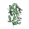
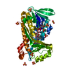
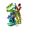
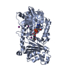
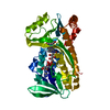
- PDB-2riv: Crystal structure of the reactive loop cleaved human Thyroxine Bi... -
+ Open data

- Basic information
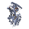
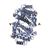
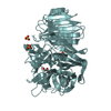
| Entry | Database: PDB / ID: 2riv | ||||||
|---|---|---|---|---|---|---|---|
| Title | Crystal structure of the reactive loop cleaved human Thyroxine Binding Globulin | ||||||
 Components Components | (Thyroxine-binding globulin) x 2 | ||||||
 Keywords Keywords | SIGNALING PROTEIN / TBG / Serpin / cleaved / thyroxine binding globulin / thyroxine / Disease mutation / Glycoprotein / Secreted | ||||||
| Function / homology |  Function and homology information Function and homology informationthyroid hormone transport / serine-type endopeptidase inhibitor activity / : / extracellular exosome / extracellular region Similarity search - Function | ||||||
| Biological species |  Homo sapiens (human) Homo sapiens (human) | ||||||
| Method |  X-RAY DIFFRACTION / SYNCHROTRON / MOLECULAR REPLACEMENT / Resolution: 1.5 Å X-RAY DIFFRACTION / SYNCHROTRON / MOLECULAR REPLACEMENT / Resolution: 1.5 Å | ||||||
 Authors Authors | Zhou, A. / Wei, Z. / Stanley, P.L.D. / Read, R.J. / Stein, P.E. / Carrell, R.W. | ||||||
 Citation Citation | Journal: J.Biol.Chem. / Year: 2011 Title: Allosteric modulation of hormone release from thyroxine and corticosteroid-binding globulins Authors: Qi, X. / Loiseau, F. / Chan, W.L. / Yan, Y. / Wei, Z. / Milroy, L.G. / Myers, R.M. / Ley, S.V. / Read, R.J. / Carrell, R.W. / Zhou, A. | ||||||
| History |
|
- Structure visualization
| Structure viewer | Molecule: MolmilJmol/JSmol |
|---|
- Downloads & links
-Download
| PDBx/mmCIF format | 2riv.cif.gz | 100.8 KB | Display | PDBx/mmCIF format |
|---|---|---|---|---|
| PDB format | pdb2riv.ent.gz | 75.2 KB | Display | PDB format |
| PDBx/mmJSON format | 2riv.json.gz | Tree view | PDBx/mmJSON format | |
| Others |  Other downloads Other downloads |
-Validation report
| Arichive directory | https://data.pdbj.org/pub/pdb/validation_reports/ri/2rivftp://data.pdbj.org/pub/pdb/validation_reports/ri/2riv | HTTPS FTP |
|---|
-Related structure data
| Related structure data |  2riwC  2xn3C 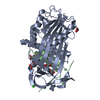 2xn5C 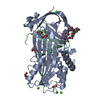 2xn6C 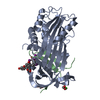 2xn7C  1qmbS C: citing same article ( S: Starting model for refinement |
|---|---|
| Similar structure data |

-Links
 PDBj
PDBj
- Assembly
| Deposited unit | 
| ||||||||
|---|---|---|---|---|---|---|---|---|---|
| 1 | 
| ||||||||
| 2 | 
| ||||||||
| 3 |
| ||||||||
| Unit cell |
| ||||||||
| Components on special symmetry positions |
|
-Components
| #1: Protein | Mass: 38399.074 Da / Num. of mol.: 1 / Fragment: N-terminal domain, UNP residues 33-375 Source method: isolated from a genetically manipulated source Details: Purified From the supernatant of the cell lysate / Source: (gene. exp.) Homo sapiens (human) / Gene: SERPINA7, TBG / Plasmid: pET16b / Production host:  | ||||||
|---|---|---|---|---|---|---|---|
| #2: Protein/peptide | Mass: 4605.383 Da / Num. of mol.: 1 Fragment: reactive loop cleaved TBG, C-teemingly domain, UNP residues 376-415 Source method: isolated from a genetically manipulated source Details: Purified From the supernatant of the cell lysate / Source: (gene. exp.) Homo sapiens (human) / Gene: SERPINA7, TBG / Plasmid: pET16b / Production host: | ||||||
| #3: Chemical |   Mass: 92.094 Da / Num. of mol.: 3 / Source method: obtained synthetically / Formula: C3H8O3 Mass: 92.094 Da / Num. of mol.: 3 / Source method: obtained synthetically / Formula: C3H8O3#4: Chemical |   Mass: 96.063 Da / Num. of mol.: 2 / Source method: obtained synthetically / Formula: SO4 Mass: 96.063 Da / Num. of mol.: 2 / Source method: obtained synthetically / Formula: SO4#5: Water | ChemComp-HOH / |  Mass: 18.015 Da / Num. of mol.: 428 / Source method: isolated from a natural source / Formula: H2O Mass: 18.015 Da / Num. of mol.: 428 / Source method: isolated from a natural source / Formula: H2OSequence details | THE REACTIVE LOOP P10-P1' CLEAVED CONFORMATION OF TBG WAS MUTATED TO GAMFLEAIPRS. THE FIRST ...THE REACTIVE LOOP P10-P1' CLEAVED CONFORMATI | |
-Experimental details
-Experiment
| Experiment | Method: X-RAY DIFFRACTION / Number of used crystals: 1 |
|---|
- Sample preparation
| Crystal | Density Matthews: 2.4 Å3/Da / Density % sol: 48.75 % |
|---|---|
| Crystal grow | Temperature: 298 K / Method: evaporation / pH: 7.4 Details: 20% PEG 3350, 0.2M sodium sulphate, pH 7.4, EVAPORATION, temperature 298K |
-Data collection
| Diffraction | Mean temperature: 100 K |
|---|---|
| Diffraction source | Source: SYNCHROTRON / Site: SRS  / Beamline: PX14.1 / Wavelength: 1.5419 Å / Beamline: PX14.1 / Wavelength: 1.5419 Å |
| Detector | Type: ADSC QUANTUM 4 / Detector: CCD / Date: May 22, 2007 / Details: mirrors |
| Radiation | Monochromator: GRAPHITE / Protocol: SINGLE WAVELENGTH / Monochromatic (M) / Laue (L): M / Scattering type: x-ray |
| Radiation wavelength | Wavelength: 1.5419 Å / Relative weight: 1 |
| Reflection | Resolution: 1.5→29.5 Å / Num. all: 64019 / Num. obs: 63059 / % possible obs: 98.5 % / Observed criterion σ(F): 2 / Observed criterion σ(I): 2 / Redundancy: 6.8 % / Rmerge(I) obs: 0.041 / Net I/σ(I): 28.9 |
| Reflection shell | Resolution: 1.5→1.539 Å / Redundancy: 4.5 % / Rmerge(I) obs: 0.222 / Mean I/σ(I) obs: 7.5 / Num. unique all: 3930 / % possible all: 84.8 |
- Processing
| Software |
| ||||||||||||||||||||||||||||||||||||||||||||||||||||||||||||||||||||||||||||||||||||||||||||||||||||||||||||||||||||||||||||||||||||||||||||||||||||||||||||||||||||||||||
|---|---|---|---|---|---|---|---|---|---|---|---|---|---|---|---|---|---|---|---|---|---|---|---|---|---|---|---|---|---|---|---|---|---|---|---|---|---|---|---|---|---|---|---|---|---|---|---|---|---|---|---|---|---|---|---|---|---|---|---|---|---|---|---|---|---|---|---|---|---|---|---|---|---|---|---|---|---|---|---|---|---|---|---|---|---|---|---|---|---|---|---|---|---|---|---|---|---|---|---|---|---|---|---|---|---|---|---|---|---|---|---|---|---|---|---|---|---|---|---|---|---|---|---|---|---|---|---|---|---|---|---|---|---|---|---|---|---|---|---|---|---|---|---|---|---|---|---|---|---|---|---|---|---|---|---|---|---|---|---|---|---|---|---|---|---|---|---|---|---|---|---|
| Refinement | Method to determine structure: MOLECULAR REPLACEMENT Starting model: PDB ENTRY 1QMB Resolution: 1.5→29.45 Å / Cor.coef. Fo:Fc: 0.962 / Cor.coef. Fo:Fc free: 0.953 / SU B: 2.768 / SU ML: 0.052 / TLS residual ADP flag: LIKELY RESIDUAL / Cross valid method: THROUGHOUT / σ(F): 2 / σ(I): 7.5 / ESU R: 0.077 / ESU R Free: 0.077 / Stereochemistry target values: MAXIMUM LIKELIHOOD / Details: HYDROGENS HAVE BEEN ADDED IN THE RIDING POSITIONS
| ||||||||||||||||||||||||||||||||||||||||||||||||||||||||||||||||||||||||||||||||||||||||||||||||||||||||||||||||||||||||||||||||||||||||||||||||||||||||||||||||||||||||||
| Solvent computation | Ion probe radii: 0.8 Å / Shrinkage radii: 0.8 Å / VDW probe radii: 1.2 Å / Solvent model: MASK | ||||||||||||||||||||||||||||||||||||||||||||||||||||||||||||||||||||||||||||||||||||||||||||||||||||||||||||||||||||||||||||||||||||||||||||||||||||||||||||||||||||||||||
| Displacement parameters | Biso mean: 18.913 Å2
| ||||||||||||||||||||||||||||||||||||||||||||||||||||||||||||||||||||||||||||||||||||||||||||||||||||||||||||||||||||||||||||||||||||||||||||||||||||||||||||||||||||||||||
| Refinement step | Cycle: LAST / Resolution: 1.5→29.45 Å
| ||||||||||||||||||||||||||||||||||||||||||||||||||||||||||||||||||||||||||||||||||||||||||||||||||||||||||||||||||||||||||||||||||||||||||||||||||||||||||||||||||||||||||
| Refine LS restraints |
| ||||||||||||||||||||||||||||||||||||||||||||||||||||||||||||||||||||||||||||||||||||||||||||||||||||||||||||||||||||||||||||||||||||||||||||||||||||||||||||||||||||||||||
| LS refinement shell | Resolution: 1.5→1.539 Å / Total num. of bins used: 20
| ||||||||||||||||||||||||||||||||||||||||||||||||||||||||||||||||||||||||||||||||||||||||||||||||||||||||||||||||||||||||||||||||||||||||||||||||||||||||||||||||||||||||||
| Refinement TLS params. | Method: refined / Refine-ID: X-RAY DIFFRACTION
| ||||||||||||||||||||||||||||||||||||||||||||||||||||||||||||||||||||||||||||||||||||||||||||||||||||||||||||||||||||||||||||||||||||||||||||||||||||||||||||||||||||||||||
| Refinement TLS group |
|
