 Movie
Movie Controller

[English] 日本語
 Yorodumi
- PDB-1pxb: CRYSTAL STRUCTURES OF MUTANT PSEUDOMONAS AERUGINOSA P-HYDROXYBENZ... -
+ Open data

- Basic information
| Entry | Database: PDB / ID: 1pxb | ||||||
|---|---|---|---|---|---|---|---|
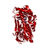
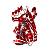
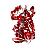
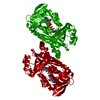
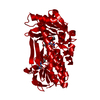
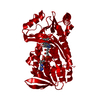
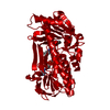
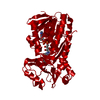
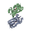
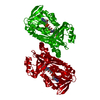
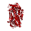
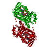
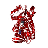
| Title | CRYSTAL STRUCTURES OF MUTANT PSEUDOMONAS AERUGINOSA P-HYDROXYBENZOATE HYDROXYLASE: THE TYR201PHE, TYR385PHE, AND ASN300ASP VARIANTS | ||||||
 Components Components | P-HYDROXYBENZOATE HYDROXYLASE | ||||||
 Keywords Keywords | OXIDOREDUCTASE | ||||||
| Function / homology |  Function and homology information Function and homology information4-hydroxybenzoate 3-monooxygenase (NADPH) activity / 4-hydroxybenzoate 3-monooxygenase / 4-hydroxybenzoate 3-monooxygenase activity / : / FAD binding / flavin adenine dinucleotide binding / oxidoreductase activity Similarity search - Function | ||||||
| Biological species |   Pseudomonas aeruginosa (bacteria) Pseudomonas aeruginosa (bacteria) | ||||||
| Method |  X-RAY DIFFRACTION / Resolution: 2.3 Å X-RAY DIFFRACTION / Resolution: 2.3 Å | ||||||
 Authors Authors | Lah, M.S. / Palfey, B.A. / Schreuder, H.A. / Ludwig, M.L. | ||||||
 Citation Citation | Journal: Biochemistry / Year: 1994 Title: Crystal structures of mutant Pseudomonas aeruginosa p-hydroxybenzoate hydroxylases: the Tyr201Phe, Tyr385Phe, and Asn300Asp variants. Authors: Lah, M.S. / Palfey, B.A. / Schreuder, H.A. / Ludwig, M.L. #1: Journal: Biochemistry / Year: 1994Title: Changes in the Catalytic Properties of P-Hydroxybenzoate Hydroxylase Caused by the Mutation Asn300Asp Authors: Palfey, B.A. / Entsch, B. / Ballou, D.P. / Massey, V. | ||||||
| History |
|
- Structure visualization
| Structure viewer | Molecule: MolmilJmol/JSmol |
|---|
- Downloads & links
-Download
| PDBx/mmCIF format | 1pxb.cif.gz | 96.2 KB | Display | PDBx/mmCIF format |
|---|---|---|---|---|
| PDB format | pdb1pxb.ent.gz | 72.6 KB | Display | PDB format |
| PDBx/mmJSON format | 1pxb.json.gz | Tree view | PDBx/mmJSON format | |
| Others |  Other downloads Other downloads |
-Validation report
| Arichive directory | https://data.pdbj.org/pub/pdb/validation_reports/px/1pxbftp://data.pdbj.org/pub/pdb/validation_reports/px/1pxb | HTTPS FTP |
|---|
-Related structure data


-Links
 PDBj
PDBj- Assembly
| Deposited unit |
| ||||||||
|---|---|---|---|---|---|---|---|---|---|
| 1 |
| ||||||||
| Unit cell |
| ||||||||
| Atom site foot note | 1: CIS PROLINE - PRO 275 |
-Components
| #1: Protein | Mass: 44366.547 Da / Num. of mol.: 1 Source method: isolated from a genetically manipulated source Source: (gene. exp.) Pseudomonas aeruginosa (bacteria)References: UniProt: P20586, 4-hydroxybenzoate 3-monooxygenase |
|---|---|
| #2: Chemical | ChemComp-FAD /   Mass: 785.550 Da / Num. of mol.: 1 / Source method: obtained synthetically / Formula: C27H33N9O15P2 / Comment: FAD*YM Mass: 785.550 Da / Num. of mol.: 1 / Source method: obtained synthetically / Formula: C27H33N9O15P2 / Comment: FAD*YM |
| #3: Chemical | ChemComp-PHB /   Mass: 138.121 Da / Num. of mol.: 1 / Source method: obtained synthetically / Formula: C7H6O3 Mass: 138.121 Da / Num. of mol.: 1 / Source method: obtained synthetically / Formula: C7H6O3 |
| #4: Water | ChemComp-HOH /  Mass: 18.015 Da / Num. of mol.: 179 / Source method: isolated from a natural source / Formula: H2O Mass: 18.015 Da / Num. of mol.: 179 / Source method: isolated from a natural source / Formula: H2O |
-Experimental details
-Experiment
| Experiment | Method: X-RAY DIFFRACTION |
|---|
- Sample preparation
| Crystal | Density Matthews: 2.6 Å3/Da / Density % sol: 52.64 % | |||||||||||||||
|---|---|---|---|---|---|---|---|---|---|---|---|---|---|---|---|---|
| Crystal grow | *PLUS Temperature: 4 ℃ / pH: 7.5 / Method: other | |||||||||||||||
| Components of the solutions | *PLUS
|
-Data collection
| Radiation | Scattering type: x-ray |
|---|---|
| Radiation wavelength | Relative weight: 1 |
| Reflection | *PLUS Highest resolution: 2.3 Å / Lowest resolution: 40 Å / Num. obs: 20499 / % possible obs: 98 % / Observed criterion σ(I): 6.63 / Redundancy: 4 % / Rmerge(I) obs: 0.123 |
- Processing
| Software |
| ||||||||||||||||||||||||||||||||||||||||||||||||||||||||||||
|---|---|---|---|---|---|---|---|---|---|---|---|---|---|---|---|---|---|---|---|---|---|---|---|---|---|---|---|---|---|---|---|---|---|---|---|---|---|---|---|---|---|---|---|---|---|---|---|---|---|---|---|---|---|---|---|---|---|---|---|---|---|
| Refinement | Resolution: 2.3→40 Å / σ(F): 0 Details: THE HYDROGEN BOND DISTANCE CUTOFF USED FOR THE TURNS IS 3.5 ANGSTROMS. THE CA(X) TO CA(X+4) DISTANCE IS LESS THAN 6.0 ANGSTROMS. THE ANGLES ARE FROM ROBSON AND GARNIER (1986) 'INTRODUCTION ...Details: THE HYDROGEN BOND DISTANCE CUTOFF USED FOR THE TURNS IS 3.5 ANGSTROMS. THE CA(X) TO CA(X+4) DISTANCE IS LESS THAN 6.0 ANGSTROMS. THE ANGLES ARE FROM ROBSON AND GARNIER (1986) 'INTRODUCTION TO PROTEINS AND PROTEIN ENGINEERING', ELSEVIER, AMSTERDAM. TYPE PHI2 PSI2 PHI3 PSI3 I -75(+-65) -30(+-40) -90(+-40) -15 TO 40 II -60(+-40) 120(+-40) 90(+-40) 0(+-40) III -75(+-65) -30(+-40) -60(+-40) -15 TO -70 I(PRIME) 75(+-65) 30(+-40) 90(+-40) -40 TO 15 II(PRIME) 60(+-40) -120(+-40) -90(+-40) 0(+-40) III(PRIME) 75(+-65) 30(+-40) 60(+-40) 15 TO 70
| ||||||||||||||||||||||||||||||||||||||||||||||||||||||||||||
| Refinement step | Cycle: LAST / Resolution: 2.3→40 Å
| ||||||||||||||||||||||||||||||||||||||||||||||||||||||||||||
| Refine LS restraints |
|
