 Movie
Movie Controller


+ Open data

- Basic information
| Entry | Database: PDB / ID: 1k0i | ||||||
|---|---|---|---|---|---|---|---|
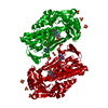
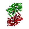
| Title | Pseudomonas aeruginosa phbh R220Q in complex with 100mM PHB | ||||||
 Components Components | P-HYDROXYBENZOATE HYDROXYLASE | ||||||
 Keywords Keywords | HYDROLASE / phbh / FAD / p-OHB | ||||||
| Function / homology |  Function and homology information Function and homology information4-hydroxybenzoate 3-monooxygenase (NADPH) activity / 4-hydroxybenzoate 3-monooxygenase / 4-hydroxybenzoate 3-monooxygenase activity / : / FAD binding / flavin adenine dinucleotide binding / oxidoreductase activity Similarity search - Function | ||||||
| Biological species |   Pseudomonas aeruginosa (bacteria) Pseudomonas aeruginosa (bacteria) | ||||||
| Method |  X-RAY DIFFRACTION / MOLECULAR REPLACEMENT / Resolution: 1.8 Å X-RAY DIFFRACTION / MOLECULAR REPLACEMENT / Resolution: 1.8 Å | ||||||
 Authors Authors | Wang, J. / Ortiz-Maldonado, M. / Entsch, B. / Ballou, D. / Gatti, D.L. | ||||||
 Citation Citation | Journal: Proc.Natl.Acad.Sci.USA / Year: 2002 Title: Protein and ligand dynamics in 4-hydroxybenzoate hydroxylase. Authors: Wang, J. / Ortiz-Maldonado, M. / Entsch, B. / Massey, V. / Ballou, D. / Gatti, D.L. | ||||||
| History |
|
- Structure visualization
| Structure viewer | Molecule: MolmilJmol/JSmol |
|---|
- Downloads & links
-Download
| PDBx/mmCIF format | 1k0i.cif.gz | 102.8 KB | Display | PDBx/mmCIF format |
|---|---|---|---|---|
| PDB format | pdb1k0i.ent.gz | 76.9 KB | Display | PDB format |
| PDBx/mmJSON format | 1k0i.json.gz | Tree view | PDBx/mmJSON format | |
| Others |  Other downloads Other downloads |
-Validation report
| Arichive directory | https://data.pdbj.org/pub/pdb/validation_reports/k0/1k0iftp://data.pdbj.org/pub/pdb/validation_reports/k0/1k0i | HTTPS FTP |
|---|
-Related structure data
| Related structure data | 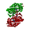 1k0jC  1k0lC 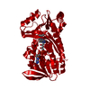 1iuwS S: Starting model for refinement C: citing same article ( |
|---|---|
| Similar structure data |


-Links
 PDBj
PDBj
- Assembly
| Deposited unit | 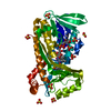
| ||||||||
|---|---|---|---|---|---|---|---|---|---|
| 1 | 
| ||||||||
| 2 | 
| ||||||||
| Unit cell |
| ||||||||
| Components on special symmetry positions |
| ||||||||
| Details | The enzyme is a dimer in solution. But only one monomer is present in the asymmetric unit |
-Components
-Protein , 1 types, 1 molecules A
| #1: Protein | Mass: 44353.484 Da / Num. of mol.: 1 / Mutation: R220Q Source method: isolated from a genetically manipulated source Source: (gene. exp.) Pseudomonas aeruginosa (bacteria) / Plasmid: bl21 / Production host: References: UniProt: P20586, 4-hydroxybenzoate 3-monooxygenase |
|---|
-Non-polymers , 5 types, 268 molecules 


| #2: Chemical | ChemComp-SO4 /  Mass: 96.063 Da / Num. of mol.: 8 / Source method: obtained synthetically / Formula: SO4 Mass: 96.063 Da / Num. of mol.: 8 / Source method: obtained synthetically / Formula: SO4#3: Chemical | ChemComp-SO3 / |  Mass: 80.063 Da / Num. of mol.: 1 / Source method: obtained synthetically / Formula: SO3 Mass: 80.063 Da / Num. of mol.: 1 / Source method: obtained synthetically / Formula: SO3#4: Chemical | ChemComp-FAD / |  Mass: 785.550 Da / Num. of mol.: 1 / Source method: obtained synthetically / Formula: C27H33N9O15P2 / Comment: FAD*YM Mass: 785.550 Da / Num. of mol.: 1 / Source method: obtained synthetically / Formula: C27H33N9O15P2 / Comment: FAD*YM#5: Chemical |  Mass: 138.121 Da / Num. of mol.: 2 / Source method: obtained synthetically / Formula: C7H6O3 Mass: 138.121 Da / Num. of mol.: 2 / Source method: obtained synthetically / Formula: C7H6O3#6: Water | ChemComp-HOH / | Mass: 18.015 Da / Num. of mol.: 256 / Source method: isolated from a natural source / Formula: H2O |
|---|
-Experimental details
-Experiment
| Experiment | Method: X-RAY DIFFRACTION / Number of used crystals: 1 |
|---|
- Sample preparation
| Crystal | Density Matthews: 2.54 Å3/Da / Density % sol: 51.66 % | ||||||||||||||||||||||||||||||||||||||||||||||||
|---|---|---|---|---|---|---|---|---|---|---|---|---|---|---|---|---|---|---|---|---|---|---|---|---|---|---|---|---|---|---|---|---|---|---|---|---|---|---|---|---|---|---|---|---|---|---|---|---|---|
| Crystal grow | Temperature: 303 K / Method: vapor diffusion, hanging drop / pH: 7 Details: 100mM potassium phosphate, 0.05 mM glutathion, 30mM sodium sulfite 0.02mM FAD, 450mM ammonium sulfate, pH 7.0, VAPOR DIFFUSION, HANGING DROP, temperature 303K | ||||||||||||||||||||||||||||||||||||||||||||||||
| Crystal grow | *PLUS Temperature: 30 ℃ | ||||||||||||||||||||||||||||||||||||||||||||||||
| Components of the solutions | *PLUS
|
-Data collection
| Diffraction | Mean temperature: 100 K |
|---|---|
| Diffraction source | Source: ROTATING ANODE / Type: RIGAKU RU200 / Wavelength: 1.5418 Å |
| Detector | Type: RIGAKU RAXIS IV / Detector: IMAGE PLATE / Date: May 20, 2001 / Details: mirrors |
| Radiation | Protocol: SINGLE WAVELENGTH / Monochromatic (M) / Laue (L): M / Scattering type: x-ray |
| Radiation wavelength | Wavelength: 1.5418 Å / Relative weight: 1 |
| Reflection | Resolution: 1.8→22.02 Å / Num. all: 38363 / Num. obs: 38363 / % possible obs: 90.6 % / Observed criterion σ(F): 0 / Observed criterion σ(I): 0 / Redundancy: 13.8 % / Biso Wilson estimate: 32.6 Å2 / Rmerge(I) obs: 0.081 |
| Reflection shell | Resolution: 1.8→1.91 Å / Rmerge(I) obs: 0.471 / Num. unique all: 3306 / % possible all: 53 |
| Reflection | *PLUS Highest resolution: 1.8 Å / Lowest resolution: 22 Å / Num. measured all: 557623 |
- Processing
| Software |
| |||||||||||||||||||||||||
|---|---|---|---|---|---|---|---|---|---|---|---|---|---|---|---|---|---|---|---|---|---|---|---|---|---|---|
| Refinement | Method to determine structure: MOLECULAR REPLACEMENT Starting model: PDB ENTRY 1IUW Resolution: 1.8→22.02 Å / Isotropic thermal model: anisotropic / Cross valid method: THROUGHOUT / σ(F): 0 / σ(I): 0 / Stereochemistry target values: Engh & Huber
| |||||||||||||||||||||||||
| Displacement parameters | Biso mean: 31.9 Å2
| |||||||||||||||||||||||||
| Refine analyze |
| |||||||||||||||||||||||||
| Refinement step | Cycle: LAST / Resolution: 1.8→22.02 Å
| |||||||||||||||||||||||||
| Refine LS restraints |
| |||||||||||||||||||||||||
| Software | *PLUS Name: CNS / Version: 1 / Classification: refinement | |||||||||||||||||||||||||
| Refinement | *PLUS Highest resolution: 1.8 Å / Lowest resolution: 22 Å / σ(F): 0 / % reflection Rfree: 10 % / Rfactor obs: 0.207 / Rfactor Rfree: 0.24 | |||||||||||||||||||||||||
| Solvent computation | *PLUS | |||||||||||||||||||||||||
| Displacement parameters | *PLUS Biso mean: 31.9 Å2 | |||||||||||||||||||||||||
| Refine LS restraints | *PLUS
|
