 Movie
Movie Controller


+ Open data

- Basic information
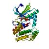
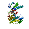
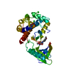
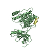
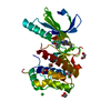
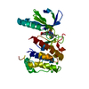
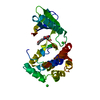
| Entry | Database: PDB / ID: 2xk7 | ||||||
|---|---|---|---|---|---|---|---|
| Title | Structure of Nek2 bound to aminopyrazine compound 23 | ||||||
 Components Components | SERINE/THREONINE-PROTEIN KINASE NEK2 | ||||||
 Keywords Keywords | TRANSFERASE / CENTROSOME / MITOSIS | ||||||
| Function / homology |  Function and homology information Function and homology informationnegative regulation of centriole-centriole cohesion / centrosome separation / regulation of attachment of spindle microtubules to kinetochore / regulation of mitotic centrosome separation / regulation of mitotic nuclear division / positive regulation of telomere maintenance / mitotic spindle assembly / blastocyst development / intercellular bridge / spindle assembly ...negative regulation of centriole-centriole cohesion / centrosome separation / regulation of attachment of spindle microtubules to kinetochore / regulation of mitotic centrosome separation / regulation of mitotic nuclear division / positive regulation of telomere maintenance / mitotic spindle assembly / blastocyst development / intercellular bridge / spindle assembly / Loss of Nlp from mitotic centrosomes / Loss of proteins required for interphase microtubule organization from the centrosome / Recruitment of mitotic centrosome proteins and complexes / Recruitment of NuMA to mitotic centrosomes / Anchoring of the basal body to the plasma membrane / AURKA Activation by TPX2 / APC-Cdc20 mediated degradation of Nek2A / condensed nuclear chromosome / meiotic cell cycle / chromosome segregation / kinetochore / spindle pole / Regulation of PLK1 Activity at G2/M Transition / mitotic cell cycle / midbody / protein phosphatase binding / microtubule / protein kinase activity / non-specific serine/threonine protein kinase / cilium / ciliary basal body / protein serine kinase activity / cell division / protein serine/threonine kinase activity / centrosome / nucleolus / protein-containing complex / nucleoplasm / ATP binding / metal ion binding / nucleus / plasma membrane / cytoplasm / cytosol Similarity search - Function | ||||||
| Biological species |  HOMO SAPIENS (human) HOMO SAPIENS (human) | ||||||
| Method |  X-RAY DIFFRACTION / SYNCHROTRON / MOLECULAR REPLACEMENT / Resolution: 1.992 Å X-RAY DIFFRACTION / SYNCHROTRON / MOLECULAR REPLACEMENT / Resolution: 1.992 Å | ||||||
 Authors Authors | Mas-Droux, C. / Bayliss, R. | ||||||
 Citation Citation | Journal: J.Med.Chem. / Year: 2010 Title: Aminopyrazine Inhibitors Binding to an Unusual Inactive Conformation of the Mitotic Kinase Nek2: Sar and Structural Characterization. Authors: Whelligan, D.K. / Solanki, S. / Taylor, D. / Thomson, D.W. / Cheung, K.M. / Boxall, K. / Mas-Droux, C. / Barillari, C. / Burns, S. / Grummitt, C.G. / Collins, I. / Van Montfort, R.L. / ...Authors: Whelligan, D.K. / Solanki, S. / Taylor, D. / Thomson, D.W. / Cheung, K.M. / Boxall, K. / Mas-Droux, C. / Barillari, C. / Burns, S. / Grummitt, C.G. / Collins, I. / Van Montfort, R.L. / Aherne, G.W. / Bayliss, R. / Hoelder, S. | ||||||
| History |
|
- Structure visualization
| Structure viewer | Molecule: MolmilJmol/JSmol |
|---|
- Downloads & links
-Download
| PDBx/mmCIF format | 2xk7.cif.gz | 121.3 KB | Display | PDBx/mmCIF format |
|---|---|---|---|---|
| PDB format | pdb2xk7.ent.gz | 93.1 KB | Display | PDB format |
| PDBx/mmJSON format | 2xk7.json.gz | Tree view | PDBx/mmJSON format | |
| Others |  Other downloads Other downloads |
-Validation report
| Arichive directory | https://data.pdbj.org/pub/pdb/validation_reports/xk/2xk7ftp://data.pdbj.org/pub/pdb/validation_reports/xk/2xk7 | HTTPS FTP |
|---|
-Related structure data
| Related structure data |  2xk3C  2xk4C 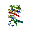 2xk6C  2xk8C  2xkcC  2xkdC  2xkeC  2xkfC  2wqoS C: citing same article ( S: Starting model for refinement |
|---|---|
| Similar structure data |


-Links
 PDBj
PDBj

- Assembly
| Deposited unit | 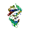
| ||||||||
|---|---|---|---|---|---|---|---|---|---|
| 1 |
| ||||||||
| Unit cell |
|
-Components
| #1: Protein | Mass: 32662.479 Da / Num. of mol.: 1 / Fragment: CATALYTIC DOMAIN, RESIDUES 1-271 Source method: isolated from a genetically manipulated source Source: (gene. exp.) HOMO SAPIENS (human) / Production host:  References: UniProt: P51955, non-specific serine/threonine protein kinase | ||
|---|---|---|---|
| #2: Chemical | ChemComp-30E / (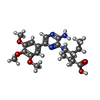  Mass: 416.471 Da / Num. of mol.: 1 / Source method: obtained synthetically / Formula: C21H28N4O5 Mass: 416.471 Da / Num. of mol.: 1 / Source method: obtained synthetically / Formula: C21H28N4O5 | ||
| #3: Chemical | ChemComp-CL /   Mass: 35.453 Da / Num. of mol.: 6 / Source method: obtained synthetically / Formula: Cl Mass: 35.453 Da / Num. of mol.: 6 / Source method: obtained synthetically / Formula: Cl#4: Water | ChemComp-HOH / |  Mass: 18.015 Da / Num. of mol.: 189 / Source method: isolated from a natural source / Formula: H2O Mass: 18.015 Da / Num. of mol.: 189 / Source method: isolated from a natural source / Formula: H2O |
-Experimental details
-Experiment
| Experiment | Method: X-RAY DIFFRACTION |
|---|
- Sample preparation
| Crystal | Density Matthews: 2.58 Å3/Da / Density % sol: 52.37 % / Description: NONE |
|---|
-Data collection
| Diffraction | Mean temperature: 100 K |
|---|---|
| Diffraction source | Source: SYNCHROTRON / Site: Diamond  / Beamline: I03 / Wavelength: 0.9763 / Beamline: I03 / Wavelength: 0.9763 |
| Detector | Type: ADSC CCD / Detector: CCD |
| Radiation | Protocol: SINGLE WAVELENGTH / Monochromatic (M) / Laue (L): M / Scattering type: x-ray |
| Radiation wavelength | Wavelength: 0.9763 Å / Relative weight: 1 |
| Reflection | Resolution: 1.99→50.37 Å / Num. obs: 22841 / % possible obs: 99.6 % / Observed criterion σ(I): 6 / Redundancy: 3.6 % / Biso Wilson estimate: 24.96 Å2 / Rmerge(I) obs: 0.069 / Net I/σ(I): 12.1 |
| Reflection shell | Resolution: 1.99→2.1 Å / Redundancy: 3.6 % / Rmerge(I) obs: 0.374 / Mean I/σ(I) obs: 3.4 / % possible all: 99.4 |
- Processing
| Software |
| |||||||||||||||||||||||||||||||||||||||||||||||||||||||||||||||||||||||||||||||||||||||||||||||||||||||||||||||||||||||||||||
|---|---|---|---|---|---|---|---|---|---|---|---|---|---|---|---|---|---|---|---|---|---|---|---|---|---|---|---|---|---|---|---|---|---|---|---|---|---|---|---|---|---|---|---|---|---|---|---|---|---|---|---|---|---|---|---|---|---|---|---|---|---|---|---|---|---|---|---|---|---|---|---|---|---|---|---|---|---|---|---|---|---|---|---|---|---|---|---|---|---|---|---|---|---|---|---|---|---|---|---|---|---|---|---|---|---|---|---|---|---|---|---|---|---|---|---|---|---|---|---|---|---|---|---|---|---|---|
| Refinement | Method to determine structure: MOLECULAR REPLACEMENT Starting model: PDB ENTRY 2WQO Resolution: 1.992→36.636 Å / SU ML: 0.25 / σ(F): 0.01 / Phase error: 21.32 / Stereochemistry target values: ML
| |||||||||||||||||||||||||||||||||||||||||||||||||||||||||||||||||||||||||||||||||||||||||||||||||||||||||||||||||||||||||||||
| Solvent computation | Shrinkage radii: 0.9 Å / VDW probe radii: 1.11 Å / Solvent model: FLAT BULK SOLVENT MODEL / Bsol: 60.773 Å2 / ksol: 0.333 e/Å3 | |||||||||||||||||||||||||||||||||||||||||||||||||||||||||||||||||||||||||||||||||||||||||||||||||||||||||||||||||||||||||||||
| Displacement parameters |
| |||||||||||||||||||||||||||||||||||||||||||||||||||||||||||||||||||||||||||||||||||||||||||||||||||||||||||||||||||||||||||||
| Refinement step | Cycle: LAST / Resolution: 1.992→36.636 Å
| |||||||||||||||||||||||||||||||||||||||||||||||||||||||||||||||||||||||||||||||||||||||||||||||||||||||||||||||||||||||||||||
| Refine LS restraints |
| |||||||||||||||||||||||||||||||||||||||||||||||||||||||||||||||||||||||||||||||||||||||||||||||||||||||||||||||||||||||||||||
| LS refinement shell |
| |||||||||||||||||||||||||||||||||||||||||||||||||||||||||||||||||||||||||||||||||||||||||||||||||||||||||||||||||||||||||||||
| Refinement TLS params. | Method: refined / Refine-ID: X-RAY DIFFRACTION
| |||||||||||||||||||||||||||||||||||||||||||||||||||||||||||||||||||||||||||||||||||||||||||||||||||||||||||||||||||||||||||||
| Refinement TLS group |
|
