Mass: 18.015 Da / Num. of mol.: 172 / Source method: isolated from a natural source / Formula: H2O
Has protein modification
Y
Sequence details
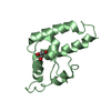
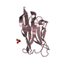
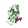
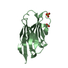
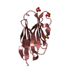
MUTATIONS DONE V5Q, A11V, G24A, A61V, A69D, S80Y.
-
Experimental details
-
Experiment
Experiment
Method: X-RAY DIFFRACTION / Number of used crystals: 1
-
Sample preparation
Crystal
Density Matthews: 2.87 Å3/Da / Density % sol: 57.2 % / Description: NONE
Crystal grow
pH: 6 Details: 5 MG/ML OF D7 PROTEIN IN A SOLUTION OF 20MM HEPES PH 7, 0.1 M NACL MIXED WITH AN EQUAL VOLUME OF 100MM SODIUM CACODYLATE PH 6, 20 MM MAGNESIUM ACETATE, 1.7M AMMONIUM SULFATE AND 19 % GLYCEROL WELL CONDITION
Resolution: 1.5→24.027 Å / SU ML: 0.17 / σ(F): 0.08 / Phase error: 15.88 / Stereochemistry target values: ML Details: DISORDERED REGIONS FOR THE CDR3 LOOP FROM RESIDUE 98 TO RESIDUE 100C.
Rfactor
Num. reflection
% reflection
Rfree
0.1935
1135
5.1 %
Rwork
0.1657
-
-
obs
0.167
22364
92.93 %
Solvent computation
Shrinkage radii: 0.9 Å / VDW probe radii: 1.11 Å / Solvent model: FLAT BULK SOLVENT MODEL / Bsol: 51.488 Å2 / ksol: 0.357 e/Å3
Displacement parameters
Baniso -1
Baniso -2
Baniso -3
1-
-1.2255 Å2
0 Å2
0 Å2
2-
-
-0.4192 Å2
0 Å2
3-
-
-
1.6447 Å2
Refinement step
Cycle: LAST / Resolution: 1.5→24.027 Å
Protein
Nucleic acid
Ligand
Solvent
Total
Num. atoms
977
0
20
172
1169
Refine LS restraints
Refine-ID
Type
Dev ideal
Number
X-RAY DIFFRACTION
f_bond_d
0.006
1199
X-RAY DIFFRACTION
f_angle_d
1.065
1652
X-RAY DIFFRACTION
f_dihedral_angle_d
16.816
434
X-RAY DIFFRACTION
f_chiral_restr
0.071
167
X-RAY DIFFRACTION
f_plane_restr
0.005
225
LS refinement shell
Resolution (Å)
Rfactor Rfree
Num. reflection Rfree
Rfactor Rwork
Num. reflection Rwork
Refine-ID
% reflection obs (%)
1.5001-1.5684
0.2226
94
0.1812
1816
X-RAY DIFFRACTION
65
1.5684-1.6511
0.1996
125
0.1617
2311
X-RAY DIFFRACTION
82
1.6511-1.7545
0.1929
149
0.1629
2737
X-RAY DIFFRACTION
98
1.7545-1.8899
0.2099
162
0.1627
2788
X-RAY DIFFRACTION
99
1.8899-2.08
0.1765
168
0.1558
2816
X-RAY DIFFRACTION
99
2.08-2.3807
0.1773
147
0.1591
2836
X-RAY DIFFRACTION
100
2.3807-2.9985
0.2215
150
0.1662
2881
X-RAY DIFFRACTION
100
2.9985-24.0298
0.1616
140
0.1618
3044
X-RAY DIFFRACTION
100
+
About Yorodumi
-
News
-
Feb 9, 2022. New format data for meta-information of EMDB entries
New format data for meta-information of EMDB entries
Version 3 of the EMDB header file is now the official format.
The previous official version 1.9 will be removed from the archive.
In the structure databanks used in Yorodumi, some data are registered as the other names, "COVID-19 virus" and "2019-nCoV". Here are the details of the virus and the list of structure data.
Jan 31, 2019. EMDB accession codes are about to change! (news from PDBe EMDB page)
EMDB accession codes are about to change! (news from PDBe EMDB page)
The allocation of 4 digits for EMDB accession codes will soon come to an end. Whilst these codes will remain in use, new EMDB accession codes will include an additional digit and will expand incrementally as the available range of codes is exhausted. The current 4-digit format prefixed with “EMD-” (i.e. EMD-XXXX) will advance to a 5-digit format (i.e. EMD-XXXXX), and so on. It is currently estimated that the 4-digit codes will be depleted around Spring 2019, at which point the 5-digit format will come into force.
The EM Navigator/Yorodumi systems omit the EMD- prefix.
Related info.:Q: What is EMD? / ID/Accession-code notation in Yorodumi/EM Navigator
Yorodumi is a browser for structure data from EMDB, PDB, SASBDB, etc.
This page is also the successor to EM Navigator detail page, and also detail information page/front-end page for Omokage search.
The word "yorodu" (or yorozu) is an old Japanese word meaning "ten thousand". "mi" (miru) is to see.
Related info.:EMDB / PDB / SASBDB / Comparison of 3 databanks / Yorodumi Search / Aug 31, 2016. New EM Navigator & Yorodumi / Yorodumi Papers / Jmol/JSmol / Function and homology information / Changes in new EM Navigator and Yorodumi
 Movie
Movie Controller

 Yorodumi
 Open data
 Basic information
 Components
 Keywords
 Function and homology information
 X-RAY DIFFRACTION /
  Authors
 Citation
 Structure visualization
 Downloads & links
 Other downloads
 PDBj
PDBj

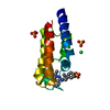
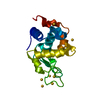
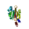
 Assembly
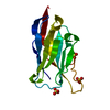


 Mass: 96.063 Da / Num. of mol.: 4 / Source method: obtained synthetically / Formula: SO4
 Mass: 18.015 Da / Num. of mol.: 172 / Source method: isolated from a natural source / Formula: H2O
 Sample preparation
 / Beamline: BM14 / Wavelength: 0.974
  Processing