Mass: 18.015 Da / Num. of mol.: 38 / Source method: isolated from a natural source / Formula: H2O
-
Details
Has protein modification
Y
Nonpolymer details
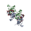
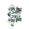
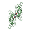
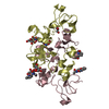
GYV, DBB (RESIDUES 1170-1173 CHAINS A, B, C, D): CANNOT BE ASSIGNED SEPARATE CHAIN IDS FOR THE ...GYV, DBB (RESIDUES 1170-1173 CHAINS A, B, C, D): CANNOT BE ASSIGNED SEPARATE CHAIN IDS FOR THE CYCLIC LIGAND. IN ABSENCE OF BACKBONE DENSITY, IT IS NOT POSSIBLE TO IDENTIFY THEIR SPECIFIC POSITIONS IN THE CYCLIC LIGAND SINCE THEY MIGHT BE ANY OF THE THREE EQUIVALENT DBB-GYV ADDUCTS.
-
Experimental details
-
Experiment
Experiment
Method: X-RAY DIFFRACTION / Number of used crystals: 1
-
Sample preparation
Crystal
Density Matthews: 2.83 Å3/Da / Density % sol: 57 % / Description: NONE
In the structure databanks used in Yorodumi, some data are registered as the other names, "COVID-19 virus" and "2019-nCoV". Here are the details of the virus and the list of structure data.
Jan 31, 2019. EMDB accession codes are about to change! (news from PDBe EMDB page)
EMDB accession codes are about to change! (news from PDBe EMDB page)
The allocation of 4 digits for EMDB accession codes will soon come to an end. Whilst these codes will remain in use, new EMDB accession codes will include an additional digit and will expand incrementally as the available range of codes is exhausted. The current 4-digit format prefixed with “EMD-” (i.e. EMD-XXXX) will advance to a 5-digit format (i.e. EMD-XXXXX), and so on. It is currently estimated that the 4-digit codes will be depleted around Spring 2019, at which point the 5-digit format will come into force.
The EM Navigator/Yorodumi systems omit the EMD- prefix.
Related info.:Q: What is EMD? / ID/Accession-code notation in Yorodumi/EM Navigator
Yorodumi is a browser for structure data from EMDB, PDB, SASBDB, etc.
This page is also the successor to EM Navigator detail page, and also detail information page/front-end page for Omokage search.
The word "yorodu" (or yorozu) is an old Japanese word meaning "ten thousand". "mi" (miru) is to see.
Related info.:EMDB / PDB / SASBDB / Comparison of 3 databanks / Yorodumi Search / Aug 31, 2016. New EM Navigator & Yorodumi / Yorodumi Papers / Jmol/JSmol / Function and homology information / Changes in new EM Navigator and Yorodumi
 Movie
Movie Controller

 Yorodumi
 Open data
 Basic information
 Components
 Keywords
 Function and homology information
 X-RAY DIFFRACTION /
  Authors
 Citation
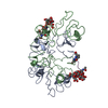
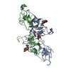
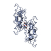
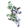
 Structure visualization
 Downloads & links
 Other downloads


 PDBj
PDBj Assembly
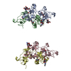

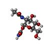
 Type: D-saccharide / Mass: 264.233 Da / Num. of mol.: 11
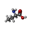


 Type: D-peptide linking / Mass: 103.120 Da / Num. of mol.: 7 / Source method: obtained synthetically / Formula: C4H9NO2
 Mass: 92.094 Da / Num. of mol.: 3 / Source method: obtained synthetically / Formula: C3H8O3
 Sample preparation
 / Beamline: ID23-1 / Wavelength: 1
  Processing