 Movie
Movie Controller

[English] 日本語
 Yorodumi
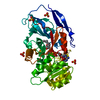
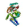
- PDB-1k7v: Crystal Structure Analysis of crosslinked-WGA3/GlcNAcbeta1,6Galbe... -
+ Open data

- Basic information
| Entry | Database: PDB / ID: 1k7v | ||||||||||||
|---|---|---|---|---|---|---|---|---|---|---|---|---|---|
| Title | Crystal Structure Analysis of crosslinked-WGA3/GlcNAcbeta1,6Galbeta1,4Glc | ||||||||||||
 Components Components | agglutinin isolectin 3 | ||||||||||||
 Keywords Keywords | SUGAR BINDING PROTEIN / Hevein-type fold | ||||||||||||
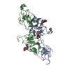
| Function / homology |  Function and homology information Function and homology information | ||||||||||||
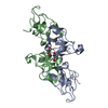
| Biological species |  | ||||||||||||
| Method |  X-RAY DIFFRACTION / MOLECULAR REPLACEMENT / Resolution: 2.2 Å X-RAY DIFFRACTION / MOLECULAR REPLACEMENT / Resolution: 2.2 Å | ||||||||||||
 Authors Authors | Muraki, M. / Ishimura, M. / Harata, K. | ||||||||||||
 Citation Citation | Journal: Biochim.Biophys.Acta / Year: 2002 Title: Interactions of wheat-germ agglutinin with GlcNAc beta 1,6Gal sequence Authors: Muraki, M. / Ishimura, M. / Harata, K. #1: Journal: Acta Crystallogr.,Sect.D / Year: 1995Title: X-ray structure of wheat germ agglutinin isolectin 3 Authors: Harata, K. / Nagahora, H. / Jigami, Y. #2: Journal: J.Mol.Biol. / Year: 2000Title: Crystal structures of Urtica dioica agglutinin and its complex with tri-N-acetylchitotriose Authors: Harata, K. / Muraki, M. #3: Journal: Protein Eng. / Year: 2000Title: Chemically prepared hevein domains: effect of C-terminal truncation and the mutagenesis of aromatic residues on the affinity for chitin Authors: Muraki, M. / Morii, H. / Harata, K. | ||||||||||||
| History |
|
- Structure visualization
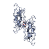
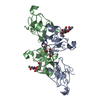
| Structure viewer | Molecule: MolmilJmol/JSmol |
|---|
- Downloads & links
-Download
| PDBx/mmCIF format | 1k7v.cif.gz | 78 KB | Display | PDBx/mmCIF format |
|---|---|---|---|---|
| PDB format | pdb1k7v.ent.gz | 57.1 KB | Display | PDB format |
| PDBx/mmJSON format | 1k7v.json.gz | Tree view | PDBx/mmJSON format | |
| Others |  Other downloads Other downloads |
-Validation report
| Arichive directory | https://data.pdbj.org/pub/pdb/validation_reports/k7/1k7vftp://data.pdbj.org/pub/pdb/validation_reports/k7/1k7v | HTTPS FTP |
|---|
-Related structure data


-Links
 PDBj
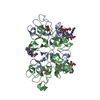
PDBj- Assembly
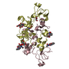
| Deposited unit | 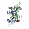
| ||||||||
|---|---|---|---|---|---|---|---|---|---|
| 1 |
| ||||||||
| 2 | 
| ||||||||
| Unit cell |
|
-Components
| #1: Protein | Mass: 18752.932 Da / Num. of mol.: 2 / Source method: isolated from a natural source / Source: (natural) #2: Polysaccharide | Source method: isolated from a genetically manipulated source #3: Water | ChemComp-HOH / |  Mass: 18.015 Da / Num. of mol.: 120 / Source method: isolated from a natural source / Formula: H2O Mass: 18.015 Da / Num. of mol.: 120 / Source method: isolated from a natural source / Formula: H2OHas protein modification | Y | |
|---|
-Experimental details
-Experiment
| Experiment | Method: X-RAY DIFFRACTION / Number of used crystals: 1 |
|---|
- Sample preparation
| Crystal | Density Matthews: 2.31 Å3/Da / Density % sol: 46.77 % | |||||||||||||||||||||||||||||||||||
|---|---|---|---|---|---|---|---|---|---|---|---|---|---|---|---|---|---|---|---|---|---|---|---|---|---|---|---|---|---|---|---|---|---|---|---|---|
| Crystal grow | Temperature: 298 K / Method: vapor diffusion, sitting drop / pH: 4.9 Details: sodium acetate, calcium chloride, Ethanol, pH 4.9, VAPOR DIFFUSION, SITTING DROP, temperature 298.0K | |||||||||||||||||||||||||||||||||||
| Crystal grow | *PLUS Method: unknownDetails: Harata, K., (1995) Acta Crystallogr., Sect.D, 51, 1013. | |||||||||||||||||||||||||||||||||||
| Components of the solutions | *PLUS
|
-Data collection
| Diffraction source | Source: ROTATING ANODE / Type: ENRAF-NONIUS FR571 / Wavelength: 1.5418 Å |
|---|---|
| Detector | Type: ENRAF-NONIUS FAST / Detector: AREA DETECTOR / Date: Dec 31, 2000 |
| Radiation | Monochromator: GRAPHITE / Protocol: SINGLE WAVELENGTH / Monochromatic (M) / Laue (L): M / Scattering type: x-ray |
| Radiation wavelength | Wavelength: 1.5418 Å / Relative weight: 1 |
| Reflection | Resolution: 2.2→23.5 Å / Num. obs: 33443 / Observed criterion σ(I): 0 / Redundancy: 2.07 % / Rmerge(I) obs: 0.102 |
| Reflection shell | Resolution: 2.2→2.24 Å / Rmerge(I) obs: 0.204 |
| Reflection | *PLUS Highest resolution: 2.2 Å / Num. obs: 16716 / % possible obs: 93.2 % / Num. measured all: 33443 |
- Processing
| Software |
| ||||||||||||||||||||
|---|---|---|---|---|---|---|---|---|---|---|---|---|---|---|---|---|---|---|---|---|---|
| Refinement | Method to determine structure: MOLECULAR REPLACEMENT Starting model: Native WGA3 Resolution: 2.2→8 Å / σ(F): 2 / Details: Average B-value are for protein atoms
| ||||||||||||||||||||
| Displacement parameters | Biso mean: 19.13 Å2 | ||||||||||||||||||||
| Refine analyze | Luzzati coordinate error obs: 0.25 Å | ||||||||||||||||||||
| Refinement step | Cycle: LAST / Resolution: 2.2→8 Å
| ||||||||||||||||||||
| Refine LS restraints |
| ||||||||||||||||||||
| Refinement | *PLUS σ(F): 2 / % reflection Rfree: 10 % / Rfactor obs: 0.232 / Rfactor Rfree: 0.31 | ||||||||||||||||||||
| Solvent computation | *PLUS | ||||||||||||||||||||
| Displacement parameters | *PLUS | ||||||||||||||||||||
| Refine LS restraints | *PLUS
|
