Monochromator: SI(111)MONOCHROMATOR / Protocol: SINGLE WAVELENGTH / Monochromatic (M) / Laue (L): M / Scattering type: x-ray
Radiation wavelength
ID
Wavelength (Å)
Relative weight
1
1.0688
1
2
0.9792
1
Reflection
Resolution: 2.3→80 Å / Num. obs: 11317 / % possible obs: 98.9 % / Observed criterion σ(I): 0 / Redundancy: 3.5 % / Biso Wilson estimate: 44.1 Å2 / Rsym value: 0.08 / Net I/σ(I): 11.9
Reflection shell
Resolution: 2.31→2.37 Å / Redundancy: 3.5 % / Mean I/σ(I) obs: 2.5 / Rsym value: 0.56 / % possible all: 95.7
-
Processing
Software
Name
Version
Classification
REFMAC
5.5.0072
refinement
XDS
datareduction
XSCALE
datascaling
autoSHARP
phasing
Refinement
Method to determine structure: SAD Starting model: NONE Resolution: 2.31→56.08 Å / Cor.coef. Fo:Fc: 0.933 / Cor.coef. Fo:Fc free: 0.882 / SU B: 5.699 / SU ML: 0.14 / Cross valid method: THROUGHOUT / ESU R: 0.206 / ESU R Free: 0.191 / Stereochemistry target values: MAXIMUM LIKELIHOOD Details: HYDROGENS HAVE BEEN ADDED IN THE RIDING POSITIONS. U VALUES REFINED INDIVIDUALLY. RESIDUES A575-A582, B532-B534, B577-B582, C532-C535 ARE DISORDERED.
Rfactor
Num. reflection
% reflection
Selection details
Rfree
0.25232
608
5.4 %
RANDOM
Rwork
0.20793
-
-
-
obs
0.21007
10707
98.92 %
-
Solvent computation
Ion probe radii: 0.8 Å / Shrinkage radii: 0.8 Å / VDW probe radii: 1.4 Å / Solvent model: MASK
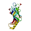
 Movie
Movie Controller


 Open data
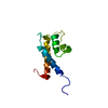
 Basic information
 Components
 Keywords
 Function and homology information
 HOMO SAPIENS (human)
 X-RAY DIFFRACTION /
  Authors
 Citation
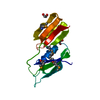
 Structure visualization
 Downloads & links
 Other downloads
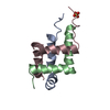


 PDBj
PDBj


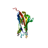
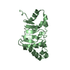
 Assembly

 Mass: 18.015 Da / Num. of mol.: 54 / Source method: isolated from a natural source / Formula: H2O
 Sample preparation
 / Beamline: X10SA / Wavelength: 1.0688, 0.9792
 Processing