 Movie
Movie Controller

[English] 日本語
 Yorodumi
- PDB-2wx4: Asymmetric trimer of the Drosophila melanogaster DCP1 C-terminal ... -
+ Open data

- Basic information
| Entry | Database: PDB / ID: 2wx4 | ||||||
|---|---|---|---|---|---|---|---|
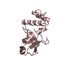
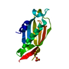
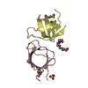
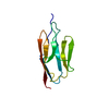
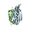
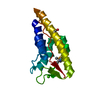
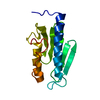
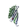
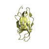
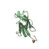
| Title | Asymmetric trimer of the Drosophila melanogaster DCP1 C-terminal domain | ||||||
 Components Components | DECAPPING PROTEIN 1 | ||||||
 Keywords Keywords | STRUCTURAL PROTEIN / ASYMMETRIC ASSEMBLY / TRIMERIZATION MODULE / MRNA DECAPPING / P-BODY COMPONENT | ||||||
| Function / homology |  Function and homology information Function and homology informationButyrate Response Factor 1 (BRF1) binds and destabilizes mRNA / Tristetraprolin (TTP, ZFP36) binds and destabilizes mRNA / mRNA decay by 5' to 3' exoribonuclease / regulation of cytoplasmic mRNA processing body assembly / pole plasm / deadenylation-independent decapping of nuclear-transcribed mRNA / pole plasm oskar mRNA localization / positive regulation of mRNA catabolic process / deadenylation-dependent decapping of nuclear-transcribed mRNA / miRNA-mediated post-transcriptional gene silencing ...Butyrate Response Factor 1 (BRF1) binds and destabilizes mRNA / Tristetraprolin (TTP, ZFP36) binds and destabilizes mRNA / mRNA decay by 5' to 3' exoribonuclease / regulation of cytoplasmic mRNA processing body assembly / pole plasm / deadenylation-independent decapping of nuclear-transcribed mRNA / pole plasm oskar mRNA localization / positive regulation of mRNA catabolic process / deadenylation-dependent decapping of nuclear-transcribed mRNA / miRNA-mediated post-transcriptional gene silencing / P granule / nuclear-transcribed mRNA catabolic process, deadenylation-dependent decay / nuclear-transcribed mRNA catabolic process / P-body / enzyme activator activity / mRNA processing / cytoplasmic stress granule / mRNA binding / cytoplasm Similarity search - Function | ||||||
| Biological species |  | ||||||
| Method |  X-RAY DIFFRACTION / SYNCHROTRON / MOLECULAR REPLACEMENT / Resolution: 2.8 Å X-RAY DIFFRACTION / SYNCHROTRON / MOLECULAR REPLACEMENT / Resolution: 2.8 Å | ||||||
 Authors Authors | Tritschler, F. / Weichenrieder, O. | ||||||
 Citation Citation | Journal: Proc.Natl.Acad.Sci.USA / Year: 2009 Title: Dcp1 Forms Asymmetric Trimers to Assemble Into Active Mrna Decapping Complexes in Metazoa. Authors: Tritschler, F. / Braun, J.E. / Motz, C. / Igreja, C. / Haas, G. / Truffault, V. / Izaurralde, E. / Weichenrieder, O. | ||||||
| History |
|
- Structure visualization
| Structure viewer | Molecule: MolmilJmol/JSmol |
|---|
- Downloads & links
-Download
| PDBx/mmCIF format | 2wx4.cif.gz | 62.3 KB | Display | PDBx/mmCIF format |
|---|---|---|---|---|
| PDB format | pdb2wx4.ent.gz | 48.3 KB | Display | PDB format |
| PDBx/mmJSON format | 2wx4.json.gz | Tree view | PDBx/mmJSON format | |
| Others |  Other downloads Other downloads |
-Validation report
| Arichive directory | https://data.pdbj.org/pub/pdb/validation_reports/wx/2wx4ftp://data.pdbj.org/pub/pdb/validation_reports/wx/2wx4 | HTTPS FTP |
|---|
-Related structure data
| Related structure data | 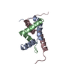 2wx3SC S: Starting model for refinement C: citing same article ( |
|---|---|
| Similar structure data |
-Links
 PDBj
PDBj
- Assembly
| Deposited unit | 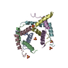
| ||||||||
|---|---|---|---|---|---|---|---|---|---|
| 1 | 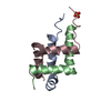
| ||||||||
| 2 | 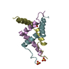
| ||||||||
| Unit cell |
|
-Components
| #1: Protein/peptide | Mass: 5215.934 Da / Num. of mol.: 6 / Fragment: TRIMERIZATION DOMAIN, RESIDUES 328-366 Source method: isolated from a genetically manipulated source Details: EC6.1.1.- IN UNIPROT DISPUTED BY AUTHOR / Source: (gene. exp.)  #2: Chemical | ChemComp-SO4 /   Mass: 96.063 Da / Num. of mol.: 5 / Source method: obtained synthetically / Formula: SO4 Mass: 96.063 Da / Num. of mol.: 5 / Source method: obtained synthetically / Formula: SO4#3: Water | ChemComp-HOH / |  Mass: 18.015 Da / Num. of mol.: 54 / Source method: isolated from a natural source / Formula: H2O Mass: 18.015 Da / Num. of mol.: 54 / Source method: isolated from a natural source / Formula: H2OSequence details | N-TERMINAL CLONING TAG - GPHMADL | |
|---|
-Experimental details
-Experiment
| Experiment | Method: X-RAY DIFFRACTION / Number of used crystals: 1 |
|---|
- Sample preparation
| Crystal | Density Matthews: 4.5 Å3/Da / Density % sol: 73 % / Description: NONE |
|---|---|
| Crystal grow | pH: 6.5 Details: 100 MM MES (PH6.5), 1.2 M AMMONIUM SULFATE, 5% 1,4-DIOXANE |
-Data collection
| Diffraction | Mean temperature: 100 K |
|---|---|
| Diffraction source | Source: SYNCHROTRON / Site: SLS  / Beamline: X10SA / Wavelength: 1.0643 / Beamline: X10SA / Wavelength: 1.0643 |
| Detector | Type: MARRESEARCH / Detector: CCD / Date: Apr 12, 2009 / Details: MIRRORS |
| Radiation | Monochromator: SI(111)MONOCHROMATOR / Protocol: SINGLE WAVELENGTH / Monochromatic (M) / Laue (L): M / Scattering type: x-ray |
| Radiation wavelength | Wavelength: 1.0643 Å / Relative weight: 1 |
| Reflection | Resolution: 2.8→50 Å / Num. obs: 14564 / % possible obs: 98.2 % / Observed criterion σ(I): 0 / Redundancy: 4.8 % / Biso Wilson estimate: 55.2 Å2 / Rsym value: 0.11 / Net I/σ(I): 13 |
| Reflection shell | Resolution: 2.8→2.87 Å / Redundancy: 4.4 % / Mean I/σ(I) obs: 2 / Rsym value: 0.8 / % possible all: 96 |
- Processing
| Software |
| ||||||||||||||||||||||||||||||||||||||||||||||||||||||||||||||||||||||||||||||||||||||||||||||||||||||||||||||||||||||||||||||||||||||||||||||||||||||||||||||||||||||||||||||||||||||
|---|---|---|---|---|---|---|---|---|---|---|---|---|---|---|---|---|---|---|---|---|---|---|---|---|---|---|---|---|---|---|---|---|---|---|---|---|---|---|---|---|---|---|---|---|---|---|---|---|---|---|---|---|---|---|---|---|---|---|---|---|---|---|---|---|---|---|---|---|---|---|---|---|---|---|---|---|---|---|---|---|---|---|---|---|---|---|---|---|---|---|---|---|---|---|---|---|---|---|---|---|---|---|---|---|---|---|---|---|---|---|---|---|---|---|---|---|---|---|---|---|---|---|---|---|---|---|---|---|---|---|---|---|---|---|---|---|---|---|---|---|---|---|---|---|---|---|---|---|---|---|---|---|---|---|---|---|---|---|---|---|---|---|---|---|---|---|---|---|---|---|---|---|---|---|---|---|---|---|---|---|---|---|---|
| Refinement | Method to determine structure: MOLECULAR REPLACEMENT Starting model: PDB ENTRY 2WX3 TRUNCATED POLY-ALA MODEL Resolution: 2.8→48.79 Å / Cor.coef. Fo:Fc: 0.923 / Cor.coef. Fo:Fc free: 0.879 / SU B: 10.537 / SU ML: 0.212 / Cross valid method: THROUGHOUT / ESU R: 0.391 / ESU R Free: 0.3 / Stereochemistry target values: MAXIMUM LIKELIHOOD Details: HYDROGENS HAVE BEEN ADDED IN THE RIDING POSITIONS. U VALUES REFINED INDIVIDUALLY. RESIDUES A359-A366,B321-B325,C364-C366,D366,E321-E322,E366,F321 ARE DISORDERED
| ||||||||||||||||||||||||||||||||||||||||||||||||||||||||||||||||||||||||||||||||||||||||||||||||||||||||||||||||||||||||||||||||||||||||||||||||||||||||||||||||||||||||||||||||||||||
| Solvent computation | Ion probe radii: 0.8 Å / Shrinkage radii: 0.8 Å / VDW probe radii: 1.4 Å / Solvent model: MASK | ||||||||||||||||||||||||||||||||||||||||||||||||||||||||||||||||||||||||||||||||||||||||||||||||||||||||||||||||||||||||||||||||||||||||||||||||||||||||||||||||||||||||||||||||||||||
| Displacement parameters | Biso mean: 43.18 Å2
| ||||||||||||||||||||||||||||||||||||||||||||||||||||||||||||||||||||||||||||||||||||||||||||||||||||||||||||||||||||||||||||||||||||||||||||||||||||||||||||||||||||||||||||||||||||||
| Refinement step | Cycle: LAST / Resolution: 2.8→48.79 Å
| ||||||||||||||||||||||||||||||||||||||||||||||||||||||||||||||||||||||||||||||||||||||||||||||||||||||||||||||||||||||||||||||||||||||||||||||||||||||||||||||||||||||||||||||||||||||
| Refine LS restraints |
|