 Movie
Movie Controller

[English] 日本語
 Yorodumi
- PDB-2ww2: Structure of the Family GH92 Inverting Mannosidase BT2199 from Ba... -
+ Open data

- Basic information
| Entry | Database: PDB / ID: 2ww2 | ||||||
|---|---|---|---|---|---|---|---|
| Title | Structure of the Family GH92 Inverting Mannosidase BT2199 from Bacteroides thetaiotaomicron VPI-5482 | ||||||
 Components Components | ALPHA-1,2-MANNOSIDASE | ||||||
 Keywords Keywords | HYDROLASE / GLYCOSIDE HYDROLASE FAMILY 92 / BT2199 | ||||||
| Function / homology |  Function and homology information Function and homology informationpeptide-N4-(N-acetyl-beta-glucosaminyl)asparagine amidase activity / glycoprotein catabolic process / carbohydrate binding / carbohydrate metabolic process / cytosol Similarity search - Function | ||||||
| Biological species |  BACTEROIDES THETAIOTAOMICRON (bacteria) BACTEROIDES THETAIOTAOMICRON (bacteria) | ||||||
| Method |  X-RAY DIFFRACTION / SYNCHROTRON / MOLECULAR REPLACEMENT / Resolution: 1.9 Å X-RAY DIFFRACTION / SYNCHROTRON / MOLECULAR REPLACEMENT / Resolution: 1.9 Å | ||||||
 Authors Authors | Suits, M.D.L. / Zhu, Y. / Thompson, A. / Gilbert, H.J. / Davies, G.J. | ||||||
 Citation Citation | Journal: Nat.Chem.Biol. / Year: 2010 Title: Mechanistic Insights Into a Ca2+-Dependent Family of A-Mannosidases in a Human Gut Symbiont. Authors: Zhu, Y. / Suits, M.D.L. / Thompson, A. / Chavan, S. / Dinev, Z. / Dumon, C. / Smith, N. / Moremen, K.W. / Xiang, Y. / Siriwardena, A. / Williams, S.J. / Gilbert, H.J. / Davies, G.J. | ||||||
| History |
|
- Structure visualization
| Structure viewer | Molecule: MolmilJmol/JSmol |
|---|
- Downloads & links
-Download
| PDBx/mmCIF format | 2ww2.cif.gz | 485.6 KB | Display | PDBx/mmCIF format |
|---|---|---|---|---|
| PDB format | pdb2ww2.ent.gz | 394.6 KB | Display | PDB format |
| PDBx/mmJSON format | 2ww2.json.gz | Tree view | PDBx/mmJSON format | |
| Others |  Other downloads Other downloads |
-Validation report
| Arichive directory | https://data.pdbj.org/pub/pdb/validation_reports/ww/2ww2ftp://data.pdbj.org/pub/pdb/validation_reports/ww/2ww2 | HTTPS FTP |
|---|
-Related structure data
| Related structure data | 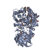 2wvxSC 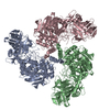 2wvyC 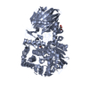 2wvzC 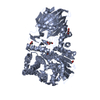 2ww0C 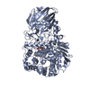 2ww1C 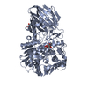 2ww3C 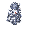 2wzsC C: citing same article ( S: Starting model for refinement |
|---|---|
| Similar structure data |


-Links
 PDBj
PDBj


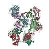
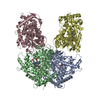
- Assembly
| Deposited unit | 
| ||||||||
|---|---|---|---|---|---|---|---|---|---|
| 1 |
| ||||||||
| Unit cell |
| ||||||||
| Components on special symmetry positions |
|
-Components
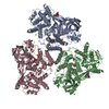
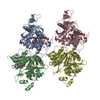
-Protein , 1 types, 3 molecules ABC
| #1: Protein | Mass: 83742.734 Da / Num. of mol.: 3 / Fragment: RESIDUES 22-758 Source method: isolated from a genetically manipulated source Source: (gene. exp.) BACTEROIDES THETAIOTAOMICRON (bacteria)Strain: VPI-5482 / Plasmid: PET 22 / Production host: |
|---|
-Non-polymers , 5 types, 2090 molecules 


| #2: Chemical | ChemComp-MPD / ( Mass: 118.174 Da / Num. of mol.: 1 / Source method: obtained synthetically / Formula: C6H14O2 / Comment: precipitant*YM Mass: 118.174 Da / Num. of mol.: 1 / Source method: obtained synthetically / Formula: C6H14O2 / Comment: precipitant*YM | ||||||
|---|---|---|---|---|---|---|---|
| #3: Chemical | ChemComp-NA /  Mass: 22.990 Da / Num. of mol.: 4 / Source method: obtained synthetically / Formula: Na Mass: 22.990 Da / Num. of mol.: 4 / Source method: obtained synthetically / Formula: Na#4: Chemical | ChemComp-GOL /  Mass: 92.094 Da / Num. of mol.: 14 / Source method: obtained synthetically / Formula: C3H8O3 Mass: 92.094 Da / Num. of mol.: 14 / Source method: obtained synthetically / Formula: C3H8O3#5: Chemical | ChemComp-SWA / |  Mass: 173.210 Da / Num. of mol.: 1 / Source method: obtained synthetically / Formula: C8H15NO3 / Comment: chemotherapy, inhibitor, alkaloid*YM Mass: 173.210 Da / Num. of mol.: 1 / Source method: obtained synthetically / Formula: C8H15NO3 / Comment: chemotherapy, inhibitor, alkaloid*YM#6: Water | ChemComp-HOH / | Mass: 18.015 Da / Num. of mol.: 2070 / Source method: isolated from a natural source / Formula: H2O |
-Experimental details
-Experiment
| Experiment | Method: X-RAY DIFFRACTION / Number of used crystals: 1 |
|---|
- Sample preparation
| Crystal | Density Matthews: 2.8 Å3/Da / Density % sol: 56 % / Description: BT3990-APO USED AS MR MODEL |
|---|---|
| Crystal grow | pH: 8 Details: 5%(V/V) MPD, 100MM HEPES PH7.5, 1.0M NA/K TARTRATE, PH 8 |
-Data collection
| Diffraction | Mean temperature: 100 K |
|---|---|
| Diffraction source | Source: SYNCHROTRON / Site: ESRF  / Beamline: ID14-1 / Wavelength: 0.9334 / Beamline: ID14-1 / Wavelength: 0.9334 |
| Detector | Type: ADSC CCD / Detector: CCD / Date: Nov 21, 2008 |
| Radiation | Protocol: SINGLE WAVELENGTH / Monochromatic (M) / Laue (L): M / Scattering type: x-ray |
| Radiation wavelength | Wavelength: 0.9334 Å / Relative weight: 1 |
| Reflection | Resolution: 1.9→36.06 Å / Num. obs: 228038 / % possible obs: 99.9 % / Observed criterion σ(I): 2 / Redundancy: 6.2 % / Rmerge(I) obs: 0.09 / Net I/σ(I): 11.2 |
| Reflection shell | Resolution: 1.9→2 Å / Redundancy: 6.1 % / Rmerge(I) obs: 0.51 / Mean I/σ(I) obs: 4.1 / % possible all: 99.9 |
- Processing
| Software |
| ||||||||||||||||||||||||||||||||||||||||||||||||||||||||||||||||||||||||||||||||||||||||||||||||||||||||||||||||||||||||||||||||||||||||||||||||||||||||||||||||||||||||||||||||||||||
|---|---|---|---|---|---|---|---|---|---|---|---|---|---|---|---|---|---|---|---|---|---|---|---|---|---|---|---|---|---|---|---|---|---|---|---|---|---|---|---|---|---|---|---|---|---|---|---|---|---|---|---|---|---|---|---|---|---|---|---|---|---|---|---|---|---|---|---|---|---|---|---|---|---|---|---|---|---|---|---|---|---|---|---|---|---|---|---|---|---|---|---|---|---|---|---|---|---|---|---|---|---|---|---|---|---|---|---|---|---|---|---|---|---|---|---|---|---|---|---|---|---|---|---|---|---|---|---|---|---|---|---|---|---|---|---|---|---|---|---|---|---|---|---|---|---|---|---|---|---|---|---|---|---|---|---|---|---|---|---|---|---|---|---|---|---|---|---|---|---|---|---|---|---|---|---|---|---|---|---|---|---|---|---|
| Refinement | Method to determine structure: MOLECULAR REPLACEMENT Starting model: PDB ENTRY 2WVX Resolution: 1.9→34.99 Å / Cor.coef. Fo:Fc: 0.967 / Cor.coef. Fo:Fc free: 0.955 / SU B: 5.655 / SU ML: 0.074 / TLS residual ADP flag: LIKELY RESIDUAL / Cross valid method: THROUGHOUT / ESU R: 0.115 / ESU R Free: 0.109 / Stereochemistry target values: MAXIMUM LIKELIHOOD / Details: HYDROGENS HAVE BEEN ADDED IN THE RIDING POSITIONS
| ||||||||||||||||||||||||||||||||||||||||||||||||||||||||||||||||||||||||||||||||||||||||||||||||||||||||||||||||||||||||||||||||||||||||||||||||||||||||||||||||||||||||||||||||||||||
| Solvent computation | Ion probe radii: 0.8 Å / Shrinkage radii: 0.8 Å / VDW probe radii: 1.2 Å / Solvent model: MASK | ||||||||||||||||||||||||||||||||||||||||||||||||||||||||||||||||||||||||||||||||||||||||||||||||||||||||||||||||||||||||||||||||||||||||||||||||||||||||||||||||||||||||||||||||||||||
| Displacement parameters | Biso mean: 16.17 Å2
| ||||||||||||||||||||||||||||||||||||||||||||||||||||||||||||||||||||||||||||||||||||||||||||||||||||||||||||||||||||||||||||||||||||||||||||||||||||||||||||||||||||||||||||||||||||||
| Refinement step | Cycle: LAST / Resolution: 1.9→34.99 Å
| ||||||||||||||||||||||||||||||||||||||||||||||||||||||||||||||||||||||||||||||||||||||||||||||||||||||||||||||||||||||||||||||||||||||||||||||||||||||||||||||||||||||||||||||||||||||
| Refine LS restraints |
|