[English] 日本語
 Yorodumi
- PDB-2wqy: Remodelling of carboxin binding to the Q-site of avian respirator... -
+ Open data
- Basic information
| Entry | Database: PDB / ID: 2wqy | |||||||||
|---|---|---|---|---|---|---|---|---|---|---|
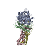
| Title | Remodelling of carboxin binding to the Q-site of avian respiratory complex II | |||||||||
 Components Components | (SUCCINATE DEHYDROGENASE ...) x 4 | |||||||||
 Keywords Keywords | OXIDOREDUCTASE / OXALOACETATE NITROPROPIONATE UBIQUINONE / RESPIRATORY CHAIN / COMPLEX II / CYTOCROME B / REDOX ENZYME / HEME PROTEIN / FLAVOPROTEIN / METAL-BINDING / MITOCHONDRION INNER MEMBRANE / IRON SULFUR PROTEIN / TRICARBOXYLIC ACID CYCLE | |||||||||
| Function / homology |  Function and homology information Function and homology informationThe tricarboxylic acid cycle / Oxidoreductases; Acting on the CH-OH group of donors; With a quinone or similar compound as acceptor / succinate metabolic process / respiratory chain complex II (succinate dehydrogenase) / mitochondrial electron transport, succinate to ubiquinone / succinate dehydrogenase (quinone) activity / succinate dehydrogenase / 3 iron, 4 sulfur cluster binding / ubiquinone binding / tricarboxylic acid cycle ...The tricarboxylic acid cycle / Oxidoreductases; Acting on the CH-OH group of donors; With a quinone or similar compound as acceptor / succinate metabolic process / respiratory chain complex II (succinate dehydrogenase) / mitochondrial electron transport, succinate to ubiquinone / succinate dehydrogenase (quinone) activity / succinate dehydrogenase / 3 iron, 4 sulfur cluster binding / ubiquinone binding / tricarboxylic acid cycle / aerobic respiration / respiratory electron transport chain / 2 iron, 2 sulfur cluster binding / mitochondrial membrane / flavin adenine dinucleotide binding / 4 iron, 4 sulfur cluster binding / electron transfer activity / mitochondrial inner membrane / heme binding / metal ion binding / membrane Similarity search - Function | |||||||||
| Biological species |  | |||||||||
| Method |  X-RAY DIFFRACTION / X-RAY DIFFRACTION /  MOLECULAR REPLACEMENT / Resolution: 2.1 Å MOLECULAR REPLACEMENT / Resolution: 2.1 Å | |||||||||
 Authors Authors | Ruprecht, J. / Iwata, S. / Cecchini, G. | |||||||||
 Citation Citation |  Journal: J.Biol.Chem. / Year: 2006 Journal: J.Biol.Chem. / Year: 2006Title: 3-Nitropropionic Acid is a Suicide Inhibitor of Mitochondrial Respiration that, Upon Oxidation by Complex II, Forms a Covalent Adduct with a Catalytic Base Arginine in the Active Site of the Enzyme. Authors: Huang, L. / Sun, G. / Cobessi, D. / Wang, A.C. / Shen, J.T. / Tung, E.Y. / Anderson, V.E. / Berry, E.A. | |||||||||
| History |
| |||||||||
| Remark 0 | THIS ENTRY 2WQY REFLECTS AN ALTERNATIVE MODELING OF THE ORIGINAL STRUCTURAL DATA (R2FBWSF) ...THIS ENTRY 2WQY REFLECTS AN ALTERNATIVE MODELING OF THE ORIGINAL STRUCTURAL DATA (R2FBWSF) DETERMINED BY AUTHORS OF THE PDB ENTRY 2FBW: L.S.HUANG,G.SUN,D.COBESSI,A.C.WANG,J.T.SHEN,E.Y.TUNG, V.E.ANDERSON,E.A.BERRY | |||||||||
| Remark 700 | THE SHEET STRUCTURE OF THIS MOLECULE IS BIFURCATED. IN ORDER TO REPRESENT THIS FEATURE IN THE ... THE SHEET STRUCTURE OF THIS MOLECULE IS BIFURCATED. IN ORDER TO REPRESENT THIS FEATURE IN THE SHEET RECORDS BELOW, TWO SHEETS ARE DEFINED. |
- Structure visualization
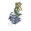
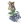
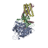
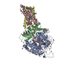
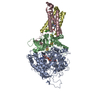
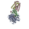
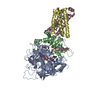
| Structure viewer | Molecule:  Molmil Molmil Jmol/JSmol Jmol/JSmol |
|---|
- Downloads & links
- Download
| PDBx/mmCIF format |  2wqy.cif.gz 2wqy.cif.gz | 497.1 KB | Display |  PDBx/mmCIF format PDBx/mmCIF format |
|---|---|---|---|---|
| PDB format |  pdb2wqy.ent.gz pdb2wqy.ent.gz | 396.5 KB | Display |  PDB format PDB format |
| PDBx/mmJSON format |  2wqy.json.gz 2wqy.json.gz | Tree view |  PDBx/mmJSON format PDBx/mmJSON format | |
| Others |  Other downloads Other downloads |
-Validation report
| Arichive directory |  https://data.pdbj.org/pub/pdb/validation_reports/wq/2wqy https://data.pdbj.org/pub/pdb/validation_reports/wq/2wqy ftp://data.pdbj.org/pub/pdb/validation_reports/wq/2wqy ftp://data.pdbj.org/pub/pdb/validation_reports/wq/2wqy | HTTPS FTP |
|---|
-Related structure data
| Related structure data |  1yq3S S: Starting model for refinement |
|---|---|
| Similar structure data |
- Links
Links
- Assembly
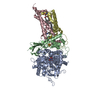
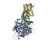
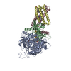
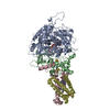
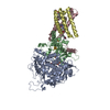
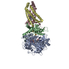
| Deposited unit | 
| ||||||||
|---|---|---|---|---|---|---|---|---|---|
| 1 | 
| ||||||||
| 2 | 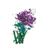
| ||||||||
| Unit cell |
|
- Components
-SUCCINATE DEHYDROGENASE ... , 4 types, 8 molecules ANBOCPDQ
| #1: Protein | Mass: 68256.922 Da / Num. of mol.: 2 / Source method: isolated from a natural source / Source: (natural)  #2: Protein | Mass: 28685.221 Da / Num. of mol.: 2 / Source method: isolated from a natural source / Source: (natural)  #3: Protein | Mass: 15419.120 Da / Num. of mol.: 2 / Source method: isolated from a natural source / Source: (natural)  #4: Protein | Mass: 10971.604 Da / Num. of mol.: 2 / Fragment: RESIDUES 55-157 / Source method: isolated from a natural source / Source: (natural)  |
|---|
-Sugars , 1 types, 2 molecules 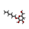
| #14: Sugar |
|---|
-Non-polymers , 13 types, 2116 molecules 




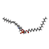


| #5: Chemical | ChemComp-UNL / Num. of mol.: 95 / Source method: obtained synthetically #6: Chemical | ChemComp-K / #7: Chemical | ChemComp-AZI / | #8: Chemical | #9: Chemical | #10: Chemical | #11: Chemical | #12: Chemical | #13: Chemical | ChemComp-GOL / #15: Chemical | #16: Chemical | #17: Chemical | #18: Water | ChemComp-HOH / | |
|---|
-Experimental details
-Experiment
| Experiment | Method:  X-RAY DIFFRACTION / Number of used crystals: 1 X-RAY DIFFRACTION / Number of used crystals: 1 |
|---|
- Sample preparation
| Crystal | Density Matthews: 3.29 Å3/Da / Density % sol: 62.3 % / Description: AUTHOR USED THE SF DATA FROM ENTRY 2FBW. |
|---|---|
| Crystal grow | Temperature: 277 K / Method: vapor diffusion, sitting drop Details: 50 G/L PEG-3350, 25 ML/L ISOPROPANOL, 15 ML/L PEG-400 0.05 M NA-HEPES, 0.01 M TRIS-HCL, 0.0005 M MNCL2, 0.0013 M MGCL2, 0.0015 M NA-AZIDE, 0.00025 M NA-EDTA, CARBOXIN, PH 7.50, VAPOR ...Details: 50 G/L PEG-3350, 25 ML/L ISOPROPANOL, 15 ML/L PEG-400 0.05 M NA-HEPES, 0.01 M TRIS-HCL, 0.0005 M MNCL2, 0.0013 M MGCL2, 0.0015 M NA-AZIDE, 0.00025 M NA-EDTA, CARBOXIN, PH 7.50, VAPOR DIFFUSION, SITTING DROP, TEMPERATURE 277K |
-Data collection
| Radiation | Protocol: SINGLE WAVELENGTH / Monochromatic (M) / Laue (L): M / Scattering type: x-ray |
|---|---|
| Radiation wavelength | Relative weight: 1 |
- Processing
| Software |
| ||||||||||||||||||||
|---|---|---|---|---|---|---|---|---|---|---|---|---|---|---|---|---|---|---|---|---|---|
| Refinement | Method to determine structure:  MOLECULAR REPLACEMENT MOLECULAR REPLACEMENTStarting model: PDB ENTRY 1YQ3 Resolution: 2.1→64.09 Å / Cor.coef. Fo:Fc: 0.942 / Cor.coef. Fo:Fc free: 0.922 / Cross valid method: THROUGHOUT / ESU R: 0.207 / ESU R Free: 0.176 / Stereochemistry target values: MAXIMUM LIKELIHOOD Details: HYDROGENS HAVE BEEN ADDED IN THE RIDING POSITIONS. U VALUES REFINED INDIVIDUALLY. STRUCTURE IS A REMODELLING OF CARBOXIN BINDING TO THE Q- SITE OF AVIAN COMPLEX II. POSITIONAL AND B-FACTOR ...Details: HYDROGENS HAVE BEEN ADDED IN THE RIDING POSITIONS. U VALUES REFINED INDIVIDUALLY. STRUCTURE IS A REMODELLING OF CARBOXIN BINDING TO THE Q- SITE OF AVIAN COMPLEX II. POSITIONAL AND B-FACTOR REFINEMENT OF CARBOXIN ONLY WAS PERFORMED. THE REST OF THE STRUCTURE IS AS MODELLED IN 2FBW.
| ||||||||||||||||||||
| Solvent computation | Ion probe radii: 0.8 Å / Shrinkage radii: 0.8 Å / VDW probe radii: 1.4 Å / Solvent model: MASK | ||||||||||||||||||||
| Displacement parameters | Biso mean: 30.499 Å2
| ||||||||||||||||||||
| Refinement step | Cycle: LAST / Resolution: 2.1→64.09 Å
| ||||||||||||||||||||
| LS refinement shell | Resolution: 2.1→2.154 Å / Total num. of bins used: 20
|
 Movie
Movie Controller






 PDBj
PDBj


