 Movie
Movie Controller

[English] 日本語
 Yorodumi
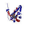
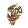
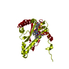
- PDB-2wqf: Crystal Structure of the Nitroreductase CinD from Lactococcus lac... -
+ Open data

- Basic information
| Entry | Database: PDB / ID: 2wqf | ||||||
|---|---|---|---|---|---|---|---|
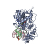
| Title | Crystal Structure of the Nitroreductase CinD from Lactococcus lactis in Complex with FMN | ||||||
 Components Components | COPPER INDUCED NITROREDUCTASE D | ||||||
 Keywords Keywords | OXIDOREDUCTASE / NITROREDUCTASE / COPR REGULATED PROTEIN | ||||||
| Function / homology |  Function and homology information Function and homology informationcellular response to oxidative stress / oxidoreductase activity / nucleotide binding / cytoplasm Similarity search - Function | ||||||
| Biological species |  LACTOCOCCUS LACTIS (lactic acid bacteria) LACTOCOCCUS LACTIS (lactic acid bacteria) | ||||||
| Method |  X-RAY DIFFRACTION / SYNCHROTRON / MOLECULAR REPLACEMENT / Resolution: 1.35 Å X-RAY DIFFRACTION / SYNCHROTRON / MOLECULAR REPLACEMENT / Resolution: 1.35 Å | ||||||
 Authors Authors | Waltersperger, S.M. / Oberholzer, A.E. / Solioz, M. / Baumann, U. | ||||||
 Citation Citation | Journal: J.Bacteriol. / Year: 2010 Title: Structure and Function of Cind (Ytjd) of Lactococcus Lactis, a Copper-Induced Nitroreductase Involved in Defense Against Oxidative Stress. Authors: Mermod, M. / Mourlane, F. / Waltersperger, S. / Oberholzer, A.E. / Baumann, U. / Solioz, M. | ||||||
| History |
|
- Structure visualization
| Structure viewer | Molecule: MolmilJmol/JSmol |
|---|
- Downloads & links
-Download
| PDBx/mmCIF format | 2wqf.cif.gz | 128.2 KB | Display | PDBx/mmCIF format |
|---|---|---|---|---|
| PDB format | pdb2wqf.ent.gz | 100.4 KB | Display | PDB format |
| PDBx/mmJSON format | 2wqf.json.gz | Tree view | PDBx/mmJSON format | |
| Others |  Other downloads Other downloads |
-Validation report
| Arichive directory | https://data.pdbj.org/pub/pdb/validation_reports/wq/2wqfftp://data.pdbj.org/pub/pdb/validation_reports/wq/2wqf | HTTPS FTP |
|---|
-Related structure data
| Related structure data | 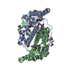 2ifaS S: Starting model for refinement |
|---|---|
| Similar structure data |


-Links
 PDBj
PDBj- Assembly
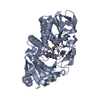
| Deposited unit | 
| ||||||||
|---|---|---|---|---|---|---|---|---|---|
| 1 | 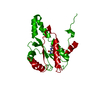
| ||||||||
| Unit cell |
| ||||||||
| Components on special symmetry positions |
|
-Components
| #1: Protein | Mass: 22562.412 Da / Num. of mol.: 1 Source method: isolated from a genetically manipulated source Source: (gene. exp.) LACTOCOCCUS LACTIS (lactic acid bacteria)Strain: IL1403 / Plasmid: PPROEX HTA / Production host: |
|---|---|
| #2: Chemical | ChemComp-FMN /   Mass: 456.344 Da / Num. of mol.: 1 / Source method: obtained synthetically / Formula: C17H21N4O9P Mass: 456.344 Da / Num. of mol.: 1 / Source method: obtained synthetically / Formula: C17H21N4O9P |
| #3: Water | ChemComp-HOH /  Mass: 18.015 Da / Num. of mol.: 296 / Source method: isolated from a natural source / Formula: H2O Mass: 18.015 Da / Num. of mol.: 296 / Source method: isolated from a natural source / Formula: H2O |
-Experimental details
-Experiment
| Experiment | Method: X-RAY DIFFRACTION |
|---|
- Sample preparation
| Crystal | Density Matthews: 2.47 Å3/Da / Density % sol: 50.2 % / Description: NONE |
|---|---|
| Crystal grow | Details: 0.2M MGCL2, 0.1M TRISHCL PH 8.0, 5% MPD (V/V), 20% PEG 3350 (W/V), 1MM FMN, 4MM DTT |
-Data collection
| Diffraction | Mean temperature: 100 K |
|---|---|
| Diffraction source | Source: SYNCHROTRON / Site: SLS  / Beamline: X06DA / Wavelength: 1 / Beamline: X06DA / Wavelength: 1 |
| Detector | Type: MARMOSAIC 225 mm CCD / Detector: CCD |
| Radiation | Protocol: SINGLE WAVELENGTH / Monochromatic (M) / Laue (L): M / Scattering type: x-ray |
| Radiation wavelength | Wavelength: 1 Å / Relative weight: 1 |
| Reflection | Resolution: 1.35→49.15 Å / Num. obs: 48865 / % possible obs: 99.2 % / Observed criterion σ(I): -3 / Redundancy: 8.9 % / Rmerge(I) obs: 0.09 / Net I/σ(I): 12.36 |
| Reflection shell | Resolution: 1.35→1.4 Å / Rmerge(I) obs: 0.58 / Mean I/σ(I) obs: 1.9 / % possible all: 97.3 |
- Processing
| Software |
| |||||||||||||||||||||||||||||||||||||||||||||||||||||||||||||||||||||||||||||
|---|---|---|---|---|---|---|---|---|---|---|---|---|---|---|---|---|---|---|---|---|---|---|---|---|---|---|---|---|---|---|---|---|---|---|---|---|---|---|---|---|---|---|---|---|---|---|---|---|---|---|---|---|---|---|---|---|---|---|---|---|---|---|---|---|---|---|---|---|---|---|---|---|---|---|---|---|---|---|
| Refinement | Method to determine structure: MOLECULAR REPLACEMENT Starting model: PDB ENTRY 2IFA Resolution: 1.35→32.134 Å / SU ML: 0 / σ(F): 2 / Phase error: 14.86 / Stereochemistry target values: ML
| |||||||||||||||||||||||||||||||||||||||||||||||||||||||||||||||||||||||||||||
| Solvent computation | Shrinkage radii: 0.9 Å / VDW probe radii: 1.11 Å / Solvent model: FLAT BULK SOLVENT MODEL / Bsol: 63.023 Å2 / ksol: 0.404 e/Å3 | |||||||||||||||||||||||||||||||||||||||||||||||||||||||||||||||||||||||||||||
| Displacement parameters |
| |||||||||||||||||||||||||||||||||||||||||||||||||||||||||||||||||||||||||||||
| Refinement step | Cycle: LAST / Resolution: 1.35→32.134 Å
| |||||||||||||||||||||||||||||||||||||||||||||||||||||||||||||||||||||||||||||
| Refine LS restraints |
| |||||||||||||||||||||||||||||||||||||||||||||||||||||||||||||||||||||||||||||
| LS refinement shell |
|
