 Movie
Movie Controller

[English] 日本語
 Yorodumi
- PDB-2ifa: Crystal Structure of the PUTATIVE NITROREDUCTASE (SMU.260) IN COM... -
+ Open data

- Basic information
| Entry | Database: PDB / ID: 2ifa | |||||||||
|---|---|---|---|---|---|---|---|---|---|---|
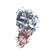
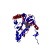
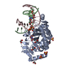
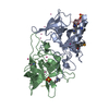
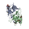
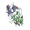
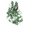
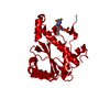
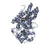
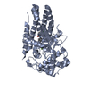
| Title | Crystal Structure of the PUTATIVE NITROREDUCTASE (SMU.260) IN COMPLEX WITH FMN FROM STREPTOCOCCUS MUTANS, NORTHEAST STRUCTURAL GENOMICS TARGET SMR5. | |||||||||
 Components Components | Hypothetical protein SMU.260 | |||||||||
 Keywords Keywords | STRUCTURAL GENOMICS / UNKNOWN FUNCTION / alpha-beta protein / PSI-2 / Protein Structure Initiative / Northeast Structural Genomics Consortium / NESG | |||||||||
| Function / homology |  Function and homology information Function and homology informationcellular response to oxidative stress / oxidoreductase activity / nucleotide binding / cytoplasm Similarity search - Function | |||||||||
| Biological species |  Streptococcus mutans (bacteria) Streptococcus mutans (bacteria) | |||||||||
| Method |  X-RAY DIFFRACTION / SYNCHROTRON / SAD / Resolution: 2.3 Å X-RAY DIFFRACTION / SYNCHROTRON / SAD / Resolution: 2.3 Å | |||||||||
 Authors Authors | Forouhar, F. / Chen, Y. / Xiao, R. / Ma, L.C. / Byler, T. / Acton, T.B. / Montelione, G.T. / Tong, L. / Hunt, J.F. / Northeast Structural Genomics Consortium (NESG) | |||||||||
| History |
|
- Structure visualization
| Structure viewer | Molecule: MolmilJmol/JSmol |
|---|
- Downloads & links
-Download
| PDBx/mmCIF format | 2ifa.cif.gz | 258.5 KB | Display | PDBx/mmCIF format |
|---|---|---|---|---|
| PDB format | pdb2ifa.ent.gz | 210.1 KB | Display | PDB format |
| PDBx/mmJSON format | 2ifa.json.gz | Tree view | PDBx/mmJSON format | |
| Others |  Other downloads Other downloads |
-Validation report
| Arichive directory | https://data.pdbj.org/pub/pdb/validation_reports/if/2ifaftp://data.pdbj.org/pub/pdb/validation_reports/if/2ifa | HTTPS FTP |
|---|
-Related structure data
| Similar structure data | |
|---|---|
| Other databases |
-Links
 PDBj
PDBj- Assembly
| Deposited unit | 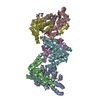
| ||||||||
|---|---|---|---|---|---|---|---|---|---|
| 1 | 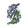
| ||||||||
| 2 | 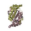
| ||||||||
| 3 | 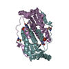
| ||||||||
| Unit cell |
|
-Components
| #1: Protein | Mass: 23662.988 Da / Num. of mol.: 6 Source method: isolated from a genetically manipulated source Source: (gene. exp.) Streptococcus mutans (bacteria) / Strain: UA159 / Gene: SMU.260 / Plasmid: pET21 / Production host: #2: Chemical | ChemComp-FMN /   Mass: 456.344 Da / Num. of mol.: 6 / Source method: obtained synthetically / Formula: C17H21N4O9P Mass: 456.344 Da / Num. of mol.: 6 / Source method: obtained synthetically / Formula: C17H21N4O9P#3: Water | ChemComp-HOH / |  Mass: 18.015 Da / Num. of mol.: 491 / Source method: isolated from a natural source / Formula: H2O Mass: 18.015 Da / Num. of mol.: 491 / Source method: isolated from a natural source / Formula: H2OHas protein modification | Y | |
|---|
-Experimental details
-Experiment
| Experiment | Method: X-RAY DIFFRACTION / Number of used crystals: 1 |
|---|
- Sample preparation
| Crystal | Density Matthews: 2.26 Å3/Da / Density % sol: 45.66 % |
|---|---|
| Crystal grow | Temperature: 293 K / Method: vapor diffusion, hanging drop / pH: 4.6 Details: 100 MM SODIUM ACETATE, 18% PEG3350, 240 MM AMMONIUM DIHYDROGEN PHOSPHATE, 10 MM FMN, AND 5 DTT., pH 4.6, VAPOR DIFFUSION, HANGING DROP, temperature 293K |
-Data collection
| Diffraction | Mean temperature: 100 K |
|---|---|
| Diffraction source | Source: SYNCHROTRON / Site: NSLS  / Beamline: X4A / Wavelength: 0.97894 Å / Beamline: X4A / Wavelength: 0.97894 Å |
| Detector | Type: ADSC QUANTUM 4 / Detector: CCD / Date: Feb 8, 2005 / Details: mirrors |
| Radiation | Monochromator: Si 111 CHANNEL / Protocol: SINGLE WAVELENGTH / Monochromatic (M) / Laue (L): M / Scattering type: x-ray |
| Radiation wavelength | Wavelength: 0.97894 Å / Relative weight: 1 |
| Reflection | Resolution: 2.3→29.03 Å / Num. all: 110600 / Num. obs: 106951 / % possible obs: 96.7 % / Observed criterion σ(F): 0 / Observed criterion σ(I): 0 / Redundancy: 4 % / Biso Wilson estimate: 11.6 Å2 / Rmerge(I) obs: 0.067 / Rsym value: 0.057 / Net I/σ(I): 17.5 |
| Reflection shell | Resolution: 2.3→2.38 Å / Redundancy: 3.7 % / Rmerge(I) obs: 0.228 / Mean I/σ(I) obs: 5.2 / Num. unique all: 10679 / Rsym value: 0.203 / % possible all: 96.7 |
- Processing
| Software |
| |||||||||||||||||||||||||||
|---|---|---|---|---|---|---|---|---|---|---|---|---|---|---|---|---|---|---|---|---|---|---|---|---|---|---|---|---|
| Refinement | Method to determine structure: SAD / Resolution: 2.3→29.03 Å / Rfactor Rfree error: 0.003 / Data cutoff high absF: 244508.83 / Data cutoff low absF: 0 / Isotropic thermal model: OVERALL / Cross valid method: THROUGHOUT / σ(F): 2 / σ(I): 2 / Stereochemistry target values: Engh & Huber
| |||||||||||||||||||||||||||
| Solvent computation | Solvent model: FLAT MODEL / Bsol: 32.4646 Å2 / ksol: 0.328468 e/Å3 | |||||||||||||||||||||||||||
| Displacement parameters | Biso mean: 28.8 Å2
| |||||||||||||||||||||||||||
| Refine analyze |
| |||||||||||||||||||||||||||
| Refinement step | Cycle: LAST / Resolution: 2.3→29.03 Å
| |||||||||||||||||||||||||||
| Refine LS restraints |
| |||||||||||||||||||||||||||
| LS refinement shell | Resolution: 2.3→2.44 Å / Rfactor Rfree error: 0.008 / Total num. of bins used: 6
|
