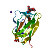
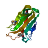
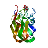
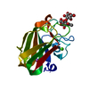
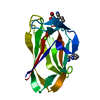
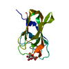
[English] 日本語
Yorodumi
- PDB-2w3j: Structure of a family 35 carbohydrate binding module from an envi... -

+
Open data


ID or keywords:

Loading...

-
Basic information

Entry
Database: PDB / ID: 2w3j
TitleStructure of a family 35 carbohydrate binding module from an environmental isolate
ComponentsCARBOHYDRATE BINDING MODULE
KeywordsSUGAR BINDING PROTEIN / SUGAR-BINDING PROTEIN / CARBOHYDRATE BINDING MODULE / FAMILY 35 / URONIC ACID SUGARS
Function / homologyGalactose-binding domain-like / Jelly Rolls / Sandwich / Mainly Beta
Function and homology information
Biological speciesUNCULTURED BACTERIUM (environmental samples)
MethodX-RAY DIFFRACTION / SYNCHROTRON / MOLECULAR REPLACEMENT / Resolution: 1.7 Å
AuthorsMontainer, C. / Flint, J. / Gloster, T.M. / Turkenburg, J.P. / Davies, G.J. / Gilbert, H.J.
CitationJournal: Proc.Natl.Acad.Sci.USA / Year: 2009
Title: Evidence that Family 35 Carbohydrate Binding Modules Display Conserved Specificity But Divergent Function.
Authors: Montanier, C. / Van Bueren, A.L. / Dumon, C. / Flint, J.E. / Correia, M.A. / Prates, J.A. / Firbank, S.J. / Lewis, R.J. / Grondin, G.G. / Ghinet, M.G. / Gloster, T.M. / Herve, C. / Knox, J.P. ...Authors: Montanier, C. / Van Bueren, A.L. / Dumon, C. / Flint, J.E. / Correia, M.A. / Prates, J.A. / Firbank, S.J. / Lewis, R.J. / Grondin, G.G. / Ghinet, M.G. / Gloster, T.M. / Herve, C. / Knox, J.P. / Talbot, B.G. / Turkenburg, J.P. / Kerovuo, J. / Brzezinski, R. / Fontes, C.M.G.A. / Davies, G.J. / Boraston, A.B. / Gilbert, H.J.
History
DepositionNov 12, 2008Deposition site: PDBE / Processing site: PDBE
Revision 1.0Jan 20, 2009Provider: repository / Type: Initial release
Revision 1.1May 8, 2011Group: Version format compliance
Revision 1.2Jul 13, 2011Group: Version format compliance
Remark 700 SHEET THE SHEET STRUCTURE OF THIS MOLECULE IS BIFURCATED. IN ORDER TO REPRESENT THIS FEATURE IN ... SHEET THE SHEET STRUCTURE OF THIS MOLECULE IS BIFURCATED. IN ORDER TO REPRESENT THIS FEATURE IN THE SHEET RECORDS BELOW, TWO SHEETS ARE DEFINED.

-
Structure visualization

Structure viewerMolecule:
MolmilJmol/JSmol

Downloads & links

-
Assembly

Deposited unit
A: CARBOHYDRATE BINDING MODULE
hetero molecules


Theoretical massNumber of molelcules
Total (without water)15,5463
Polymers15,4661
Non-polymers802
Water3,999222
1


  • Idetical with deposited unit
  • defined by author&software
TypeNameSymmetry operationNumber
identity operation1_555x,y,z1
MethodPQS
Unit cell
Length a, b, c (Å)83.943, 83.943, 79.498
Angle α, β, γ (deg.)90.00, 90.00, 120.00
Int Tables number146
Space group name H-MH3
Components on special symmetry positions
IDModelComponents
11A-2013-

HOH

21A-2044-

HOH

-
Components

#1: Protein CARBOHYDRATE BINDING MODULE


Mass: 15466.075 Da / Num. of mol.: 1
Source method: isolated from a genetically manipulated source
Source: (gene. exp.) UNCULTURED BACTERIUM (environmental samples)
Production host: ESCHERICHIA COLI (E. coli)
#2: Chemical ChemComp-CA / CALCIUM ION


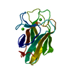
Mass: 40.078 Da / Num. of mol.: 2 / Source method: obtained synthetically / Formula: Ca
#3: Water ChemComp-HOH / water / Water


Mass: 18.015 Da / Num. of mol.: 222 / Source method: isolated from a natural source / Formula: H2O
Sequence detailsTHE FIRST RESIDUE AND FINAL 8 RESIDUES ARE INTRODUCED FROM THE EXPRESSION VECTOR.

-
Experimental details

-
Experiment

ExperimentMethod: X-RAY DIFFRACTION / Number of used crystals: 1

-
Sample preparation

CrystalDensity Matthews: 3.5 Å3/Da / Density % sol: 65 % / Description: NONE
Crystal growpH: 4.2
Details: 20% POLYETHYLENE GLYCOL 8000, 0.2M NACL, 0.25M NA CITRATE PH 4.2, 0.25M NA DIHYDROGEN PHOSPHATE PH 4.2

-
Data collection

DiffractionMean temperature: 100 K
Diffraction sourceSource: SYNCHROTRON / Site: ESRF / Beamline: ID14-1 / Wavelength: 0.9786
DetectorType: ADSC CCD / Detector: CCD
RadiationProtocol: SINGLE WAVELENGTH / Monochromatic (M) / Laue (L): M / Scattering type: x-ray
Radiation wavelengthWavelength: 0.9786 Å / Relative weight: 1
ReflectionResolution: 1.7→42 Å / Num. obs: 22914 / % possible obs: 99.7 % / Redundancy: 5.3 % / Rmerge(I) obs: 0.07 / Net I/σ(I): 15.7
Reflection shellResolution: 1.7→1.79 Å / Redundancy: 3.6 % / Rmerge(I) obs: 0.48 / Mean I/σ(I) obs: 2.5 / % possible all: 99.7

-
Processing

Software
NameVersionClassification
REFMAC5.4.0077refinement
MOSFLMdata reduction
SCALAdata scaling
AMoREphasing
RefinementMethod to determine structure: MOLECULAR REPLACEMENT / Resolution: 1.7→53.68 Å / Cor.coef. Fo:Fc: 0.97 / Cor.coef. Fo:Fc free: 0.961 / SU B: 1.667 / SU ML: 0.054 / Cross valid method: THROUGHOUT / ESU R: 0.08 / ESU R Free: 0.081 / Stereochemistry target values: MAXIMUM LIKELIHOOD / Details: HYDROGENS HAVE BEEN ADDED IN THE RIDING POSITIONS.
RfactorNum. reflection% reflectionSelection details
Rfree0.188 1177 5.1 %RANDOM
Rwork0.162 ---
obs0.163 21735 99.7 %-
Solvent computationIon probe radii: 0.8 Å / Shrinkage radii: 0.8 Å / VDW probe radii: 1.2 Å / Solvent model: MASK
Displacement parametersBiso mean: 22.86 Å2
Baniso -1Baniso -2Baniso -3
1-0.23 Å20.11 Å20 Å2
2--0.23 Å20 Å2
3----0.34 Å2
Refinement stepCycle: LAST / Resolution: 1.7→53.68 Å
ProteinNucleic acidLigandSolventTotal
Num. atoms1010 0 2 222 1234
Refine LS restraints
Refine-IDTypeDev idealDev ideal targetNumber
X-RAY DIFFRACTIONr_bond_refined_d0.0140.0221137
X-RAY DIFFRACTIONr_bond_other_d
X-RAY DIFFRACTIONr_angle_refined_deg1.3671.9251576
X-RAY DIFFRACTIONr_angle_other_deg
X-RAY DIFFRACTIONr_dihedral_angle_1_deg6.2595170
X-RAY DIFFRACTIONr_dihedral_angle_2_deg33.8822548
X-RAY DIFFRACTIONr_dihedral_angle_3_deg12.07615188
X-RAY DIFFRACTIONr_dihedral_angle_4_deg10.079156
X-RAY DIFFRACTIONr_chiral_restr0.1090.2187
X-RAY DIFFRACTIONr_gen_planes_refined0.0060.021879
X-RAY DIFFRACTIONr_gen_planes_other
X-RAY DIFFRACTIONr_nbd_refined
X-RAY DIFFRACTIONr_nbd_other
X-RAY DIFFRACTIONr_nbtor_refined
X-RAY DIFFRACTIONr_nbtor_other
X-RAY DIFFRACTIONr_xyhbond_nbd_refined
X-RAY DIFFRACTIONr_xyhbond_nbd_other
X-RAY DIFFRACTIONr_metal_ion_refined
X-RAY DIFFRACTIONr_metal_ion_other
X-RAY DIFFRACTIONr_symmetry_vdw_refined
X-RAY DIFFRACTIONr_symmetry_vdw_other
X-RAY DIFFRACTIONr_symmetry_hbond_refined
X-RAY DIFFRACTIONr_symmetry_hbond_other
X-RAY DIFFRACTIONr_symmetry_metal_ion_refined
X-RAY DIFFRACTIONr_symmetry_metal_ion_other
X-RAY DIFFRACTIONr_mcbond_it0.9121.5729
X-RAY DIFFRACTIONr_mcbond_other
X-RAY DIFFRACTIONr_mcangle_it1.65621195
X-RAY DIFFRACTIONr_mcangle_other
X-RAY DIFFRACTIONr_scbond_it2.2643408
X-RAY DIFFRACTIONr_scbond_other
X-RAY DIFFRACTIONr_scangle_it3.4274.5363
X-RAY DIFFRACTIONr_scangle_other
X-RAY DIFFRACTIONr_long_range_B_refined
X-RAY DIFFRACTIONr_long_range_B_other
X-RAY DIFFRACTIONr_rigid_bond_restr
X-RAY DIFFRACTIONr_sphericity_free
X-RAY DIFFRACTIONr_sphericity_bonded
LS refinement shellResolution: 1.7→1.74 Å / Total num. of bins used: 20 /
RfactorNum. reflection
Rfree0.371 103
Rwork0.327 1526

+
About Yorodumi

-
News

-
Feb 9, 2022. New format data for meta-information of EMDB entries

New format data for meta-information of EMDB entries

  • Version 3 of the EMDB header file is now the official format.
  • The previous official version 1.9 will be removed from the archive.

Related info.:EMDB header

External links:wwPDB to switch to version 3 of the EMDB data model

-
Aug 12, 2020. Covid-19 info

Covid-19 info

URL: https://pdbj.org/emnavi/covid19.php

New page: Covid-19 featured information page in EM Navigator.

Related info.:Covid-19 info / Mar 5, 2020. Novel coronavirus structure data

+
Mar 5, 2020. Novel coronavirus structure data

Novel coronavirus structure data

Related info.:Yorodumi Speices / Aug 12, 2020. Covid-19 info

External links:COVID-19 featured content - PDBj / Molecule of the Month (242):Coronavirus Proteases

+
Jan 31, 2019. EMDB accession codes are about to change! (news from PDBe EMDB page)

EMDB accession codes are about to change! (news from PDBe EMDB page)

  • The allocation of 4 digits for EMDB accession codes will soon come to an end. Whilst these codes will remain in use, new EMDB accession codes will include an additional digit and will expand incrementally as the available range of codes is exhausted. The current 4-digit format prefixed with “EMD-” (i.e. EMD-XXXX) will advance to a 5-digit format (i.e. EMD-XXXXX), and so on. It is currently estimated that the 4-digit codes will be depleted around Spring 2019, at which point the 5-digit format will come into force.
  • The EM Navigator/Yorodumi systems omit the EMD- prefix.

Related info.:Q: What is EMD? / ID/Accession-code notation in Yorodumi/EM Navigator

External links:EMDB Accession Codes are Changing Soon! / Contact to PDBj

+
Jul 12, 2017. Major update of PDB

Major update of PDB

  • wwPDB released updated PDB data conforming to the new PDBx/mmCIF dictionary.
  • This is a major update changing the version number from 4 to 5, and with Remediation, in which all the entries are updated.
  • In this update, many items about electron microscopy experimental information are reorganized (e.g. em_software).
  • Now, EM Navigator and Yorodumi are based on the updated data.

External links:wwPDB Remediation / Enriched Model Files Conforming to OneDep Data Standards Now Available in the PDB FTP Archive

-
Yorodumi

Thousand views of thousand structures

  • Yorodumi is a browser for structure data from EMDB, PDB, SASBDB, etc.
  • This page is also the successor to EM Navigator detail page, and also detail information page/front-end page for Omokage search.
  • The word "yorodu" (or yorozu) is an old Japanese word meaning "ten thousand". "mi" (miru) is to see.

Related info.:EMDB / PDB / SASBDB / Comparison of 3 databanks / Yorodumi Search / Aug 31, 2016. New EM Navigator & Yorodumi / Yorodumi Papers / Jmol/JSmol / Function and homology information / Changes in new EM Navigator and Yorodumi

Read more