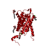
Entry Database : PDB / ID : 2w1pTitle 1.4 Angstrom crystal structure of P.pastoris aquaporin, Aqy1, in a closed conformation at pH 8.0 AQUAPORIN PIP2-7 7; Keywords / / Function / homology Function Domain/homology Component
/ / / / / / / / / Biological species KOMAGATAELLA PASTORIS (fungus)Method / / / Resolution : 1.4 Å Authors Fischer, G. / Kosinska-Eriksson, U. / Aponte-Santamaria, C. / Palmgren, M. / Geijer, C. / Hedfalk, K. / Hohmann, S. / de Groot, B.L. / Neutze, R. / Lindkvist-Petersson, K. Journal : Plos Biol. / Year : 2009Title : Crystal Structure of a Yeast Aquaporin at 1.15 A Reveals a Novel Gating MechanismAuthors : Fischer, G. / Kosinska-Eriksson, U. / Aponte-Santamaria, C. / Palmgren, M. / Geijer, C. / Hedfalk, K. / Hohmann, S. / De Groot, B.L. / Neutze, R. / Lindkvist-Petersson, K. History Deposition Oct 20, 2008 Deposition site / Processing site Revision 1.0 Jun 16, 2009 Provider / Type Revision 1.1 Apr 11, 2012 Group Database references / Derived calculations ... Database references / Derived calculations / Non-polymer description / Other / Refinement description / Source and taxonomy / Structure summary / Version format compliance Revision 1.2 Jul 29, 2020 Group Advisory / Data collection ... Advisory / Data collection / Derived calculations / Other / Structure summary Category chem_comp / entity ... chem_comp / entity / pdbx_chem_comp_identifier / pdbx_database_status / pdbx_entity_nonpoly / pdbx_unobs_or_zero_occ_atoms / struct_site / struct_site_gen Item _chem_comp.mon_nstd_flag / _chem_comp.name ... _chem_comp.mon_nstd_flag / _chem_comp.name / _chem_comp.type / _entity.pdbx_description / _pdbx_database_status.status_code_sf / _pdbx_entity_nonpoly.name Description / Provider / Type Revision 1.3 Dec 13, 2023 Group Advisory / Data collection ... Advisory / Data collection / Database references / Refinement description / Structure summary Category chem_comp / chem_comp_atom ... chem_comp / chem_comp_atom / chem_comp_bond / database_2 / pdbx_initial_refinement_model / pdbx_unobs_or_zero_occ_atoms Item / _database_2.pdbx_DOI / _database_2.pdbx_database_accession
Show all Show less
 Movie
Movie Controller

 Yorodumi
 Open data
 Basic information
 Components
 Keywords
 Function and homology information
 KOMAGATAELLA PASTORIS (fungus)
 X-RAY DIFFRACTION /
  Authors
 Citation
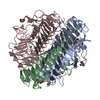
 Structure visualization
 Downloads & links
 Other downloads


 PDBj
PDBj
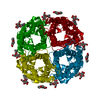
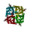
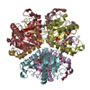
 Assembly
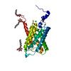


 Type: D-saccharide / Mass: 292.369 Da / Num. of mol.: 4
 Mass: 35.453 Da / Num. of mol.: 2 / Source method: obtained synthetically / Formula: Cl
 Mass: 18.015 Da / Num. of mol.: 133 / Source method: isolated from a natural source / Formula: H2O
 Sample preparation
 / Beamline: ID23-2 / Wavelength: 0.873
  Processing