 Movie
Movie Controller

[English] 日本語
 Yorodumi
- PDB-2vyu: CRYSTAL STRUCTURE OF CHOLINE BINDING PROTEIN F FROM STREPTOCOCCUS... -
+ Open data

- Basic information
| Entry | Database: PDB / ID: 2vyu | ||||||
|---|---|---|---|---|---|---|---|
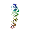
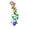
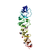
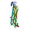
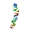
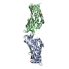
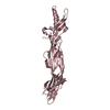
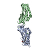
| Title | CRYSTAL STRUCTURE OF CHOLINE BINDING PROTEIN F FROM STREPTOCOCCUS PNEUMONIAE IN THE PRESENCE OF A PEPTIDOGLYCAN ANALOGUE (TETRASACCHARIDE-PENTAPEPTIDE) | ||||||
 Components Components | CHOLINE BINDING PROTEIN F | ||||||
 Keywords Keywords | CHOLINE-BINDING PROTEIN / CBPF / PEPTIDOGLYCAN / CHOLINE-BINDING-PROTEIN / LIPID-BINDING PROTEIN | ||||||
| Function / homology |  Function and homology information Function and homology informationleft handed beta-beta-3-solenoid - #20 / Cholin Binding / left handed beta-beta-3-solenoid / Choline-binding repeat / Putative cell wall binding repeat / Cell wall/choline-binding repeat / Cell wall-binding repeat profile. / Ribbon / Mainly Beta Similarity search - Domain/homology | ||||||
| Biological species |   STREPTOCOCCUS PNEUMONIAE (bacteria) STREPTOCOCCUS PNEUMONIAE (bacteria) | ||||||
| Method |  X-RAY DIFFRACTION / SYNCHROTRON / MOLECULAR REPLACEMENT / Resolution: 2.45 Å X-RAY DIFFRACTION / SYNCHROTRON / MOLECULAR REPLACEMENT / Resolution: 2.45 Å | ||||||
 Authors Authors | Perez-Dorado, I. / Molina, R. / Hermoso, J.A. / Mobashery, S. | ||||||
 Citation Citation | Journal: Embo Rep. / Year: 2009 Title: Crystal Structure of Cbpf, a Bifunctional Choline-Binding Protein and Autolysis Regulator from Streptococcus Pneumoniae. Authors: Molina, R. / Gonzalez, A. / Stelter, M. / Perez-Dorado, I. / Kahn, R. / Morales, M. / Campuzano, S. / Campillo, N.E. / Mobashery, S. / Garcia, J.L. / Garcia, P. / Hermoso, J.A. #1: Journal: Acta Crystallogr.,Sect.F / Year: 2007 Title: Crystallization and Preliminary X-Ray Diffraction Studies of Choline-Binding Protein F from Streptococcus Pneumoniae. Authors: Molina, R. / Gonzalez, A. / Moscoso, M. / Garcia, P. / Stelter, M. / Kahn, R. / Hermoso, J.A. | ||||||
| History |
|
- Structure visualization
| Structure viewer | Molecule: MolmilJmol/JSmol |
|---|
- Downloads & links
-Download
| PDBx/mmCIF format | 2vyu.cif.gz | 82.6 KB | Display | PDBx/mmCIF format |
|---|---|---|---|---|
| PDB format | pdb2vyu.ent.gz | 61.3 KB | Display | PDB format |
| PDBx/mmJSON format | 2vyu.json.gz | Tree view | PDBx/mmJSON format | |
| Others |  Other downloads Other downloads |
-Validation report
| Summary document | 2vyu_validation.pdf.gz | 441.2 KB | Display | wwPDB validaton report |
|---|---|---|---|---|
| Full document | 2vyu_full_validation.pdf.gz | 446.6 KB | Display | |
| Data in XML | 2vyu_validation.xml.gz | 16.8 KB | Display | |
| Data in CIF | 2vyu_validation.cif.gz | 24.8 KB | Display | |
| Arichive directory | https://data.pdbj.org/pub/pdb/validation_reports/vy/2vyuftp://data.pdbj.org/pub/pdb/validation_reports/vy/2vyu | HTTPS FTP |
-Related structure data
| Related structure data | 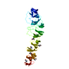 2v04C  2v05S C: citing same article ( S: Starting model for refinement |
|---|---|
| Similar structure data |


-Links
 PDBj
PDBj
- Assembly
| Deposited unit | 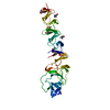
| ||||||||
|---|---|---|---|---|---|---|---|---|---|
| 1 | 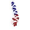
| ||||||||
| Unit cell |
|
-Components
| #1: Protein | Mass: 39383.820 Da / Num. of mol.: 1 Source method: isolated from a genetically manipulated source Source: (gene. exp.) STREPTOCOCCUS PNEUMONIAE (bacteria) / Strain: R6 / Production host: | ||
|---|---|---|---|
| #2: Chemical | ChemComp-CHT /   Mass: 104.171 Da / Num. of mol.: 7 / Source method: obtained synthetically / Formula: C5H14NO Mass: 104.171 Da / Num. of mol.: 7 / Source method: obtained synthetically / Formula: C5H14NO#3: Water | ChemComp-HOH / |  Mass: 18.015 Da / Num. of mol.: 277 / Source method: isolated from a natural source / Formula: H2O Mass: 18.015 Da / Num. of mol.: 277 / Source method: isolated from a natural source / Formula: H2O |
-Experimental details
-Experiment
| Experiment | Method: X-RAY DIFFRACTION / Number of used crystals: 1 |
|---|
- Sample preparation
| Crystal | Density Matthews: 2.78 Å3/Da / Density % sol: 55.8 % / Description: NONE |
|---|
-Data collection
| Diffraction | Mean temperature: 100 K |
|---|---|
| Diffraction source | Source: SYNCHROTRON / Site: ESRF  / Beamline: ID29 / Wavelength: 0.953746 / Beamline: ID29 / Wavelength: 0.953746 |
| Detector | Type: ADSC CCD / Detector: CCD / Date: Mar 15, 2008 |
| Radiation | Protocol: SINGLE WAVELENGTH / Monochromatic (M) / Laue (L): M / Scattering type: x-ray |
| Radiation wavelength | Wavelength: 0.953746 Å / Relative weight: 1 |
| Reflection | Resolution: 2.45→45.2 Å / Num. obs: 26724 / % possible obs: 84 % / Observed criterion σ(I): 2 / Redundancy: 5.5 % / Biso Wilson estimate: 22.8 Å2 / Rmerge(I) obs: 0.19 / Net I/σ(I): 9.7 |
| Reflection shell | Resolution: 2.45→2.69 Å / Redundancy: 5.5 % / Rmerge(I) obs: 0.56 / Mean I/σ(I) obs: 7.9 / % possible all: 87 |
- Processing
| Software |
| ||||||||||||||||||||||||||||||||||||||||||||||||||||||||||||
|---|---|---|---|---|---|---|---|---|---|---|---|---|---|---|---|---|---|---|---|---|---|---|---|---|---|---|---|---|---|---|---|---|---|---|---|---|---|---|---|---|---|---|---|---|---|---|---|---|---|---|---|---|---|---|---|---|---|---|---|---|---|
| Refinement | Method to determine structure: MOLECULAR REPLACEMENT Starting model: PDB ENTRY 2V05 Resolution: 2.45→41.84 Å / Rfactor Rfree error: 0.009 / Isotropic thermal model: RESTRAINED / Cross valid method: THROUGHOUT / σ(F): 0 Details: RESIDUES FROM 168 TO 178 ARE DISORDERED SO THEY DO NOT APPEAR IN THE MODEL.
| ||||||||||||||||||||||||||||||||||||||||||||||||||||||||||||
| Solvent computation | Solvent model: FLAT MODEL / Bsol: 48.79 Å2 / ksol: 0.38 e/Å3 | ||||||||||||||||||||||||||||||||||||||||||||||||||||||||||||
| Displacement parameters | Biso mean: 17.4 Å2
| ||||||||||||||||||||||||||||||||||||||||||||||||||||||||||||
| Refine analyze |
| ||||||||||||||||||||||||||||||||||||||||||||||||||||||||||||
| Refinement step | Cycle: LAST / Resolution: 2.45→41.84 Å
| ||||||||||||||||||||||||||||||||||||||||||||||||||||||||||||
| Refine LS restraints |
| ||||||||||||||||||||||||||||||||||||||||||||||||||||||||||||
| Refine LS restraints NCS | NCS model details: NONE | ||||||||||||||||||||||||||||||||||||||||||||||||||||||||||||
| LS refinement shell | Resolution: 2.45→2.6 Å / Rfactor Rfree error: 0.026 / Total num. of bins used: 6
|
