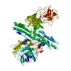
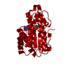
[English] 日本語
Yorodumi- PDB-2vua: Crystal Structure of the Botulinum Neurotoxin Serotype A binding ... -
+Open data
-Basic information
Entry | Database: PDB / ID: 2vua | ||||||
---|---|---|---|---|---|---|---|
Title | Crystal Structure of the Botulinum Neurotoxin Serotype A binding domain | ||||||
Components | BOTULINUM NEUROTOXIN A HEAVY CHAIN | ||||||
Keywords | HYDROLASE / GANGLIOSIDE / RECEPTOR / TOXIN / MEMBRANE / SECRETED / METAL-BINDING / METALLOPROTEASE / NEUROTOXIN / PROTEASE | ||||||
Function / homology | Function and homology information host cell junction / negative regulation of neurotransmitter secretion / bontoxilysin / host cell presynaptic membrane / host cell cytoplasmic vesicle / host cell cytosol / protein transmembrane transporter activity / metalloendopeptidase activity / toxin activity / membrane => GO:0016020 ...host cell junction / negative regulation of neurotransmitter secretion / bontoxilysin / host cell presynaptic membrane / host cell cytoplasmic vesicle / host cell cytosol / protein transmembrane transporter activity / metalloendopeptidase activity / toxin activity / membrane => GO:0016020 / host cell plasma membrane / proteolysis / zinc ion binding / extracellular region / membrane Similarity search - Function | ||||||
Biological species | CLOSTRIDIUM BOTULINUM (bacteria) | ||||||
Method | X-RAY DIFFRACTION / SYNCHROTRON / MOLECULAR REPLACEMENT / Resolution: 1.7 Å | ||||||
Authors | Stenmark, P. / Dupuy, J. / Stevens, R.C. | ||||||
Citation | Journal: Plos Pathog. / Year: 2008 Title: Crystal Structure of Botulinum Neurotoxin Type a in Complex with the Cell Surface Co-Receptor Gt1B- Insight Into the Toxin-Neuron Interaction. Authors: Stenmark, P. / Dupuy, J. / Imamura, A. / Kiso, M. / Stevens, R.C. | ||||||
History |
| ||||||
Remark 700 | SHEET THE SHEET STRUCTURE OF THIS MOLECULE IS BIFURCATED. IN ORDER TO REPRESENT THIS FEATURE IN ... SHEET THE SHEET STRUCTURE OF THIS MOLECULE IS BIFURCATED. IN ORDER TO REPRESENT THIS FEATURE IN THE SHEET RECORDS BELOW, TWO SHEETS ARE DEFINED. |
-Structure visualization
Structure viewer | Molecule: MolmilJmol/JSmol |
---|
-Downloads & links
-Download
PDBx/mmCIF format | 2vua.cif.gz | 108.7 KB | Display | PDBx/mmCIF format |
---|---|---|---|---|
PDB format | pdb2vua.ent.gz | 82.4 KB | Display | PDB format |
PDBx/mmJSON format | 2vua.json.gz | Tree view | PDBx/mmJSON format | |
Others | Other downloads |
-Validation report
Arichive directory | https://data.pdbj.org/pub/pdb/validation_reports/vu/2vua ftp://data.pdbj.org/pub/pdb/validation_reports/vu/2vua | HTTPS FTP |
---|
-Related structure data
Related structure data | 2vu9C 2nyyS C: citing same article (ref.) S: Starting model for refinement |
---|---|
Similar structure data |
-Links
-Assembly
Deposited unit |
| ||||||||
---|---|---|---|---|---|---|---|---|---|
1 |
| ||||||||
Unit cell |
|
-Components
#1: Protein | Mass: 51713.398 Da / Num. of mol.: 1 / Fragment: BINDING DOMAIN, RESIDUES 876-1296 Source method: isolated from a genetically manipulated source Source: (gene. exp.) CLOSTRIDIUM BOTULINUM (bacteria) / Strain: A1 / Plasmid: PET28A / Production host: ESCHERICHIA COLI (E. coli) / Strain (production host): BL21 References: UniProt: P10845, UniProt: P0DPI1*PLUS, bontoxilysin |
---|---|
#2: Water | ChemComp-HOH / |
-Experimental details
-Experiment
Experiment | Method: X-RAY DIFFRACTION / Number of used crystals: 1 |
---|
-Sample preparation
Crystal | Density Matthews: 2.21 Å3/Da / Density % sol: 44 % / Description: NONE |
---|---|
Crystal grow | pH: 5.2 / Details: 18% PEG3350, 0.2 M MGCL2, 0.1 M BISTRIS PH 5.2 |
-Data collection
Diffraction | Mean temperature: 100 K |
---|---|
Diffraction source | Source: SYNCHROTRON / Site: SSRL / Beamline: BL11-1 / Wavelength: 0.979 |
Detector | Type: MARRESEARCH / Detector: CCD / Date: Nov 18, 2007 |
Radiation | Protocol: SINGLE WAVELENGTH / Monochromatic (M) / Laue (L): M / Scattering type: x-ray |
Radiation wavelength | Wavelength: 0.979 Å / Relative weight: 1 |
Reflection | Resolution: 1.7→20 Å / Num. obs: 48513 / % possible obs: 99 % / Observed criterion σ(I): 0 / Redundancy: 7.8 % / Rmerge(I) obs: 0.04 / Net I/σ(I): 27 |
Reflection shell | Resolution: 1.7→1.8 Å / Redundancy: 7.5 % / Rmerge(I) obs: 0.53 / Mean I/σ(I) obs: 4.2 / % possible all: 98.4 |
-Processing
Software |
| ||||||||||||||||||||||||||||||||||||||||||||||||||||||||||||||||||||||||||||||||||||||||||||||||||||||||||||||||||||||||||||||||||||||||||||||||||||||||||||||||||||||||||||||||||||||
---|---|---|---|---|---|---|---|---|---|---|---|---|---|---|---|---|---|---|---|---|---|---|---|---|---|---|---|---|---|---|---|---|---|---|---|---|---|---|---|---|---|---|---|---|---|---|---|---|---|---|---|---|---|---|---|---|---|---|---|---|---|---|---|---|---|---|---|---|---|---|---|---|---|---|---|---|---|---|---|---|---|---|---|---|---|---|---|---|---|---|---|---|---|---|---|---|---|---|---|---|---|---|---|---|---|---|---|---|---|---|---|---|---|---|---|---|---|---|---|---|---|---|---|---|---|---|---|---|---|---|---|---|---|---|---|---|---|---|---|---|---|---|---|---|---|---|---|---|---|---|---|---|---|---|---|---|---|---|---|---|---|---|---|---|---|---|---|---|---|---|---|---|---|---|---|---|---|---|---|---|---|---|---|
Refinement | Method to determine structure: MOLECULAR REPLACEMENT Starting model: PDB ENTRY 2NYY Resolution: 1.7→19.73 Å / Cor.coef. Fo:Fc: 0.968 / Cor.coef. Fo:Fc free: 0.955 / SU B: 4.189 / SU ML: 0.07 / TLS residual ADP flag: LIKELY RESIDUAL / Cross valid method: THROUGHOUT / ESU R: 0.103 / ESU R Free: 0.102 / Stereochemistry target values: MAXIMUM LIKELIHOOD / Details: HYDROGENS HAVE BEEN ADDED IN THE RIDING POSITIONS.
| ||||||||||||||||||||||||||||||||||||||||||||||||||||||||||||||||||||||||||||||||||||||||||||||||||||||||||||||||||||||||||||||||||||||||||||||||||||||||||||||||||||||||||||||||||||||
Solvent computation | Ion probe radii: 0.8 Å / Shrinkage radii: 0.8 Å / VDW probe radii: 1.2 Å / Solvent model: BABINET MODEL WITH MASK | ||||||||||||||||||||||||||||||||||||||||||||||||||||||||||||||||||||||||||||||||||||||||||||||||||||||||||||||||||||||||||||||||||||||||||||||||||||||||||||||||||||||||||||||||||||||
Displacement parameters | Biso mean: 19.73 Å2
| ||||||||||||||||||||||||||||||||||||||||||||||||||||||||||||||||||||||||||||||||||||||||||||||||||||||||||||||||||||||||||||||||||||||||||||||||||||||||||||||||||||||||||||||||||||||
Refinement step | Cycle: LAST / Resolution: 1.7→19.73 Å
| ||||||||||||||||||||||||||||||||||||||||||||||||||||||||||||||||||||||||||||||||||||||||||||||||||||||||||||||||||||||||||||||||||||||||||||||||||||||||||||||||||||||||||||||||||||||
Refine LS restraints |
|