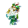
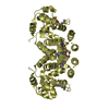
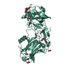
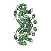
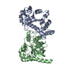
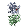
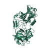
Entry Database : PDB / ID : 6f0pTitle Botulinum neurotoxin A4 Hc domain Neurotoxin type A Keywords / / / / Function / homology Function Domain/homology Component
/ / / / / / / / / / / / / / / / / / / / / / / / / / / / / / / / / / Biological species Clostridium botulinum (bacteria)Method / / / Resolution : 1.34 Å Authors Davies, J.R. / Rees, J. / Liu, S.M. / Acharya, K.R. Journal : J. Struct. Biol. / Year : 2018Title : High resolution crystal structures of Clostridium botulinum neurotoxin A3 and A4 binding domains.Authors : Davies, J.R. / Rees, J. / Liu, S.M. / Acharya, K.R. History Deposition Nov 20, 2017 Deposition site / Processing site Revision 1.0 Jan 10, 2018 Provider / Type Revision 1.1 Apr 4, 2018 Group / Database references / Category Item _citation.journal_volume / _citation.page_first ... _citation.journal_volume / _citation.page_first / _citation.page_last / _citation.year Revision 1.2 Jan 17, 2024 Group / Database references / Refinement descriptionCategory chem_comp_atom / chem_comp_bond ... chem_comp_atom / chem_comp_bond / database_2 / pdbx_initial_refinement_model Item / _database_2.pdbx_database_accessionRevision 1.3 Nov 6, 2024 Group / Category / pdbx_modification_feature
Show all Show less
 Movie
Movie Controller


 Open data
 Basic information
 Components
 Keywords
 Function and homology information
 Clostridium botulinum (bacteria)
 X-RAY DIFFRACTION /
  Authors
 Citation
 Structure visualization
 Downloads & links
 Other downloads
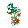


 PDBj
PDBj Assembly
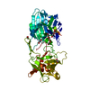


 Mass: 58.693 Da / Num. of mol.: 1 / Source method: obtained synthetically / Formula: Ni
 Mass: 106.120 Da / Num. of mol.: 2 / Source method: obtained synthetically / Formula: C4H10O3
 Mass: 62.068 Da / Num. of mol.: 5 / Source method: obtained synthetically / Formula: C2H6O2
 Mass: 209.240 Da / Num. of mol.: 1 / Source method: obtained synthetically / Formula: C8H19NO5 / Comment: pH buffer*YM
 Sample preparation
 / Beamline: I03 / Wavelength: 0.9763 Å
 Processing