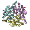
Entry Database : PDB / ID : 2vpvTitle Dimerization Domain of Mif2p PROTEIN MIF2 Keywords / / / / / / / / / Function / homology Function Domain/homology Component
/ / / / / / / / / / / / / / / / / / / Biological species SACCHAROMYCES CEREVISIAE (brewer's yeast)Method / / / Resolution : 2.7 Å Authors Cohen, R.L. / Espelin, C.W. / Sorger, P.K. / Harrison, S.C. / Simons, K.T. Journal : Mol.Biol.Cell / Year : 2008Title : Structural and Functional Dissection of Mif2P, a Conserved DNA-Binding Kinetochore Protein.Authors : Cohen, R.L. / Espelin, C.W. / De Wulf, P. / Sorger, P.K. / Harrison, S.C. / Simons, K.T. History Deposition Mar 5, 2008 Deposition site / Processing site Revision 1.0 Aug 26, 2008 Provider / Type Revision 1.1 Jun 2, 2011 Group Revision 1.2 Jul 13, 2011 Group Revision 1.3 May 8, 2019 Group Advisory / Data collection ... Advisory / Data collection / Derived calculations / Experimental preparation / Other Category database_PDB_rev / database_PDB_rev_record ... database_PDB_rev / database_PDB_rev_record / exptl_crystal_grow / pdbx_database_proc / pdbx_database_status / pdbx_struct_special_symmetry / pdbx_unobs_or_zero_occ_residues Item / _pdbx_database_status.recvd_author_approvalRevision 1.4 Nov 6, 2024 Group Advisory / Data collection ... Advisory / Data collection / Database references / Other / Structure summary Category chem_comp_atom / chem_comp_bond ... chem_comp_atom / chem_comp_bond / database_2 / pdbx_database_status / pdbx_entry_details / pdbx_modification_feature / pdbx_unobs_or_zero_occ_residues Item / _database_2.pdbx_database_accession / _pdbx_database_status.status_code_sf
Show all Show less
 Movie
Movie Controller


 Open data
 Basic information
 Components
 Keywords
 Function and homology information
 X-RAY DIFFRACTION /
  Authors
 Citation
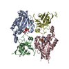
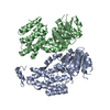
 Structure visualization
 Downloads & links
 Other downloads
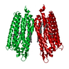


 PDBj
PDBj Assembly
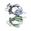


 Mass: 96.063 Da / Num. of mol.: 1 / Source method: obtained synthetically / Formula: SO4
 Mass: 18.015 Da / Num. of mol.: 27 / Source method: isolated from a natural source / Formula: H2O
 Sample preparation
 / Beamline: 8.2.2 / Wavelength: 1.5003
  Processing