 Movie
Movie Controller

[English] 日本語
 Yorodumi
- PDB-2vjv: Crystal structure of the IS608 transposase in complex with left e... -
+ Open data

- Basic information
| Entry | Database: PDB / ID: 2vjv | ||||||
|---|---|---|---|---|---|---|---|
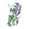
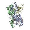
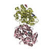
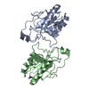
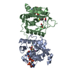
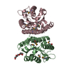
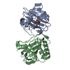
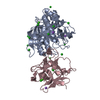
| Title | Crystal structure of the IS608 transposase in complex with left end 26-mer DNA hairpin and a 6-mer DNA representing the left end cleavage site | ||||||
 Components Components |
| ||||||
 Keywords Keywords | DNA BINDING PROTEIN / DNA-BINDING PROTEIN / PROTEIN-DNA COMPLEX / HUH MOTIF / DNA STEM LOOP / TRANSPOSITION | ||||||
| Function / homology |  Function and homology information Function and homology informationtransposase activity / DNA transposition / DNA binding / metal ion binding Similarity search - Function | ||||||
| Biological species |   HELICOBACTER PYLORI (bacteria) HELICOBACTER PYLORI (bacteria) | ||||||
| Method |  X-RAY DIFFRACTION / MOLECULAR REPLACEMENT / Resolution: 1.9 Å X-RAY DIFFRACTION / MOLECULAR REPLACEMENT / Resolution: 1.9 Å | ||||||
 Authors Authors | Barabas, O. / Ronning, D.R. / Guynet, C. / Hickman, A.B. / Ton-Hoang, B. / Chandler, M. / Dyda, F. | ||||||
 Citation Citation | Journal: Cell(Cambridge,Mass.) / Year: 2008 Title: Mechanism of is200/is605 Family DNA Transposases: Activation and Transposon-Directed Target Site Selection. Authors: Barabas, O. / Ronning, D.R. / Guynet, C. / Hickman, A.B. / Ton-Hoang, B. / Chandler, M. / Dyda, F. | ||||||
| History |
|
- Structure visualization
| Structure viewer | Molecule: MolmilJmol/JSmol |
|---|
- Downloads & links
-Download
| PDBx/mmCIF format | 2vjv.cif.gz | 107.7 KB | Display | PDBx/mmCIF format |
|---|---|---|---|---|
| PDB format | pdb2vjv.ent.gz | 78 KB | Display | PDB format |
| PDBx/mmJSON format | 2vjv.json.gz | Tree view | PDBx/mmJSON format | |
| Others |  Other downloads Other downloads |
-Validation report
| Arichive directory | https://data.pdbj.org/pub/pdb/validation_reports/vj/2vjvftp://data.pdbj.org/pub/pdb/validation_reports/vj/2vjv | HTTPS FTP |
|---|
-Related structure data
| Related structure data | 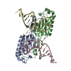 2vhgC 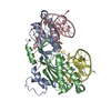 2vicC 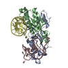 2vihSC 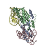 2vjuC C: citing same article ( S: Starting model for refinement |
|---|---|
| Similar structure data |


-Links
 PDBj
PDBj


- Assembly
| Deposited unit | 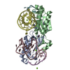
| ||||||||
|---|---|---|---|---|---|---|---|---|---|
| 1 |
| ||||||||
| Unit cell |
| ||||||||
| Noncrystallographic symmetry (NCS) | NCS oper: (Code: given Matrix: (-0.985391, 0.170267, -0.003787), Vector: |
-Components
| #1: Protein | Mass: 18392.430 Da / Num. of mol.: 2 / Fragment: RESIDUES 2-155 Source method: isolated from a genetically manipulated source Source: (gene. exp.) HELICOBACTER PYLORI (bacteria) / Strain: PECAN2A / Production host: #2: DNA chain | Mass: 8003.165 Da / Num. of mol.: 2 / Source method: obtained synthetically / Source: (synth.) HELICOBACTER PYLORI (bacteria)#3: DNA chain | Mass: 1783.216 Da / Num. of mol.: 2 / Source method: obtained synthetically / Source: (synth.) HELICOBACTER PYLORI (bacteria)#4: Chemical |   Mass: 24.305 Da / Num. of mol.: 3 / Source method: obtained synthetically / Formula: Mg Mass: 24.305 Da / Num. of mol.: 3 / Source method: obtained synthetically / Formula: Mg#5: Water | ChemComp-HOH / |  Mass: 18.015 Da / Num. of mol.: 275 / Source method: isolated from a natural source / Formula: H2O Mass: 18.015 Da / Num. of mol.: 275 / Source method: isolated from a natural source / Formula: H2OSequence details | THE ADDITION OF THE FIRST 5 RESIDUES (GSAMA) TO THE DATABASE SEQUENCE ARE THE RESULT OF CLONING ARTIFACT | |
|---|
-Experimental details
-Experiment
| Experiment | Method: X-RAY DIFFRACTION / Number of used crystals: 1 |
|---|
- Sample preparation
| Crystal | Density Matthews: 2.29 Å3/Da / Density % sol: 50.75 % / Description: NONE |
|---|---|
| Crystal grow | pH: 5 / Details: 6-10% PEG 600 AND 50 MM SODIUM CITRATE PH 5.0 |
-Data collection
| Diffraction | Mean temperature: 95 K |
|---|---|
| Diffraction source | Source: ROTATING ANODE / Type: RIGAKU MICROMAX-007 HF / Wavelength: 1.5418 |
| Detector | Type: RIGAKU IMAGE PLATE / Detector: IMAGE PLATE / Date: Jun 12, 2007 / Details: MULTILAYER FOCUSING OPTICS |
| Radiation | Monochromator: MULTILAYER FOCUSING MIRROR / Protocol: SINGLE WAVELENGTH / Monochromatic (M) / Laue (L): M / Scattering type: x-ray |
| Radiation wavelength | Wavelength: 1.5418 Å / Relative weight: 1 |
| Reflection | Resolution: 1.9→40 Å / Num. obs: 38044 / % possible obs: 89.8 % / Observed criterion σ(I): 0 / Redundancy: 3.9 % / Biso Wilson estimate: 17.3 Å2 / Rmerge(I) obs: 0.15 / Net I/σ(I): 11.3 |
| Reflection shell | Resolution: 1.9→1.95 Å / Redundancy: 1.9 % / Rmerge(I) obs: 0.27 / Mean I/σ(I) obs: 4.6 / % possible all: 64.2 |
- Processing
| Software |
| ||||||||||||||||||||||||||||||||||||||||||||||||||||||||||||||||||||||||||||||||
|---|---|---|---|---|---|---|---|---|---|---|---|---|---|---|---|---|---|---|---|---|---|---|---|---|---|---|---|---|---|---|---|---|---|---|---|---|---|---|---|---|---|---|---|---|---|---|---|---|---|---|---|---|---|---|---|---|---|---|---|---|---|---|---|---|---|---|---|---|---|---|---|---|---|---|---|---|---|---|---|---|---|
| Refinement | Method to determine structure: MOLECULAR REPLACEMENT Starting model: PDB ENTRY 2VIH Resolution: 1.9→24.61 Å / Rfactor Rfree error: 0.007 / Data cutoff high absF: 1368904.85 / Isotropic thermal model: RESTRAINED / Cross valid method: THROUGHOUT / σ(F): 0
| ||||||||||||||||||||||||||||||||||||||||||||||||||||||||||||||||||||||||||||||||
| Solvent computation | Solvent model: FLAT MODEL / Bsol: 36.2464 Å2 / ksol: 0.353019 e/Å3 | ||||||||||||||||||||||||||||||||||||||||||||||||||||||||||||||||||||||||||||||||
| Displacement parameters | Biso mean: 29.6 Å2
| ||||||||||||||||||||||||||||||||||||||||||||||||||||||||||||||||||||||||||||||||
| Refine analyze |
| ||||||||||||||||||||||||||||||||||||||||||||||||||||||||||||||||||||||||||||||||
| Refinement step | Cycle: LAST / Resolution: 1.9→24.61 Å
| ||||||||||||||||||||||||||||||||||||||||||||||||||||||||||||||||||||||||||||||||
| Refine LS restraints |
| ||||||||||||||||||||||||||||||||||||||||||||||||||||||||||||||||||||||||||||||||
| LS refinement shell | Resolution: 1.9→1.97 Å / Rfactor Rfree error: 0.036 / Total num. of bins used: 10
| ||||||||||||||||||||||||||||||||||||||||||||||||||||||||||||||||||||||||||||||||
| Xplor file |
|
