 Movie
Movie Controller

[English] 日本語
 Yorodumi
- PDB-2a6o: Crystal Structure of the ISHp608 Transposase in Complex with Stem... -
+ Open data

- Basic information
| Entry | Database: PDB / ID: 2a6o | ||||||
|---|---|---|---|---|---|---|---|
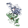
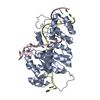
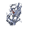
| Title | Crystal Structure of the ISHp608 Transposase in Complex with Stem-loop DNA | ||||||
 Components Components |
| ||||||
 Keywords Keywords | TRANSCRIPTION/DNA / RNA Recognition Motif / DNA Stem-loop / TRANSCRIPTION-DNA COMPLEX | ||||||
| Function / homology |  Function and homology information Function and homology informationtransposase activity / DNA transposition / DNA binding / metal ion binding Similarity search - Function | ||||||
| Biological species |   Helicobacter pylori (bacteria) Helicobacter pylori (bacteria) | ||||||
| Method |  X-RAY DIFFRACTION / MOLECULAR REPLACEMENT / Resolution: 2.6 Å X-RAY DIFFRACTION / MOLECULAR REPLACEMENT / Resolution: 2.6 Å | ||||||
 Authors Authors | Ronning, D.R. / Guynet, C. / Ton-Hoang, B. / Perez, Z.N. / Ghirlando, R. / Chandler, M. / Dyda, F. | ||||||
 Citation Citation | Journal: Mol.Cell / Year: 2005 Title: Active site sharing and subterminal hairpin recognition in a new class of DNA transposases. Authors: Ronning, D.R. / Guynet, C. / Ton-Hoang, B. / Perez, Z.N. / Ghirlando, R. / Chandler, M. / Dyda, F. | ||||||
| History |
|
- Structure visualization
| Structure viewer | Molecule: MolmilJmol/JSmol |
|---|
- Downloads & links
-Download
| PDBx/mmCIF format | 2a6o.cif.gz | 100.5 KB | Display | PDBx/mmCIF format |
|---|---|---|---|---|
| PDB format | pdb2a6o.ent.gz | 74.6 KB | Display | PDB format |
| PDBx/mmJSON format | 2a6o.json.gz | Tree view | PDBx/mmJSON format | |
| Others |  Other downloads Other downloads |
-Validation report
| Arichive directory | https://data.pdbj.org/pub/pdb/validation_reports/a6/2a6oftp://data.pdbj.org/pub/pdb/validation_reports/a6/2a6o | HTTPS FTP |
|---|
-Related structure data
| Related structure data | 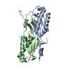 2a6mSC S: Starting model for refinement C: citing same article ( |
|---|---|
| Similar structure data |


-Links
 PDBj
PDBj


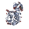
- Assembly
| Deposited unit | 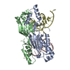
| ||||||||
|---|---|---|---|---|---|---|---|---|---|
| 1 |
| ||||||||
| Unit cell |
|
-Components
| #1: DNA chain | Mass: 6743.353 Da / Num. of mol.: 2 / Source method: obtained synthetically #2: Protein | Mass: 18106.145 Da / Num. of mol.: 2 Source method: isolated from a genetically manipulated source Source: (gene. exp.) Helicobacter pylori (bacteria) / Plasmid: pDR32 / Species (production host): Escherichia coli / Production host: #3: Water | ChemComp-HOH / |  Mass: 18.015 Da / Num. of mol.: 123 / Source method: isolated from a natural source / Formula: H2O Mass: 18.015 Da / Num. of mol.: 123 / Source method: isolated from a natural source / Formula: H2O |
|---|
-Experimental details
-Experiment
| Experiment | Method: X-RAY DIFFRACTION / Number of used crystals: 1 |
|---|
- Sample preparation
| Crystal | Density Matthews: 2.4 Å3/Da / Density % sol: 48.8 % |
|---|---|
| Crystal grow | Temperature: 293 K / Method: vapor diffusion, sitting drop / pH: 7.5 Details: PEG 3350, Sodium Tartrate, pH 7.5, VAPOR DIFFUSION, SITTING DROP, temperature 293K |
-Data collection
| Diffraction | Mean temperature: 95 K |
|---|---|
| Diffraction source | Source: ROTATING ANODE / Type: RIGAKU RU200 / Wavelength: 1.54178 Å |
| Detector | Type: RIGAKU RAXIS IV / Detector: IMAGE PLATE / Date: Nov 24, 2004 / Details: mirrors |
| Radiation | Monochromator: mirror / Protocol: SINGLE WAVELENGTH / Monochromatic (M) / Laue (L): M / Scattering type: x-ray |
| Radiation wavelength | Wavelength: 1.54178 Å / Relative weight: 1 |
| Reflection | Resolution: 2.6→50 Å / Num. obs: 14684 / % possible obs: 99.7 % / Observed criterion σ(F): 0 / Observed criterion σ(I): 0 / Redundancy: 3.6 % / Biso Wilson estimate: 36.4 Å2 / Rmerge(I) obs: 0.06 / Rsym value: 0.06 / Net I/σ(I): 14.7 |
- Processing
| Software |
| ||||||||||||||||||||||||||||||||||||
|---|---|---|---|---|---|---|---|---|---|---|---|---|---|---|---|---|---|---|---|---|---|---|---|---|---|---|---|---|---|---|---|---|---|---|---|---|---|
| Refinement | Method to determine structure: MOLECULAR REPLACEMENT Starting model: 2A6M Resolution: 2.6→44.2 Å / Rfactor Rfree error: 0.006 / Data cutoff high absF: 324112.11 / Data cutoff low absF: 0 / Isotropic thermal model: RESTRAINED / Cross valid method: THROUGHOUT / σ(F): 0 / σ(I): 0 / Stereochemistry target values: Engh & Huber
| ||||||||||||||||||||||||||||||||||||
| Solvent computation | Solvent model: FLAT MODEL / Bsol: 41.2368 Å2 / ksol: 0.371023 e/Å3 | ||||||||||||||||||||||||||||||||||||
| Displacement parameters | Biso mean: 38.5 Å2
| ||||||||||||||||||||||||||||||||||||
| Refine analyze |
| ||||||||||||||||||||||||||||||||||||
| Refinement step | Cycle: LAST / Resolution: 2.6→44.2 Å
| ||||||||||||||||||||||||||||||||||||
| Refine LS restraints |
| ||||||||||||||||||||||||||||||||||||
| LS refinement shell | Resolution: 2.6→2.76 Å / Rfactor Rfree error: 0.019 / Total num. of bins used: 6
| ||||||||||||||||||||||||||||||||||||
| Xplor file |
|
