- PDB-2vf7: Crystal structure of UvrA2 from Deinococcus radiodurans -
+
Open data
ID or keywords:
Loading...
-
Basic information
Entry
Database: PDB / ID: 2vf7
Title
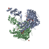
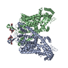
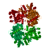
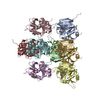
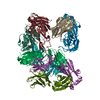
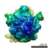
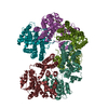
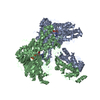
Crystal structure of UvrA2 from Deinococcus radiodurans
Components
EXCINUCLEASE ABC, SUBUNIT A.
Keywords
DNA BINDING PROTEIN / DNA-BINDING PROTEIN / NUCLEOTIDE-BINDING / ZINC-BINDING DOMAIN / SOS RESPONSE / METAL-BINDING / EXCISION NUCLEASE / ZINC-FINGER / ATP-BINDING / DNA-BINDING / DNA EXCISION / ZINC / CYTOPLASM / DNA DAMAGE / DNA REPAIR / ABC PROTEIN DNA-BINDING PROTEIN
Function / homology
Function and homology information
excinuclease repair complex / nuclease activity / nucleotide-excision repair / DNA damage response / ATP hydrolysis activity / DNA binding / zinc ion binding / ATP binding / identical protein binding / cytosol Similarity search - Function
Mass: 18.015 Da / Num. of mol.: 597 / Source method: isolated from a natural source / Formula: H2O
Compound details
ENGINEERED RESIDUE IN CHAIN A, GLN 826 TO ARG ENGINEERED RESIDUE IN CHAIN B, GLN 826 TO ARG ...ENGINEERED RESIDUE IN CHAIN A, GLN 826 TO ARG ENGINEERED RESIDUE IN CHAIN B, GLN 826 TO ARG ENGINEERED RESIDUE IN CHAIN C, GLN 826 TO ARG
-
Experimental details
-
Experiment
Experiment
Method: X-RAY DIFFRACTION / Number of used crystals: 1
-
Sample preparation
Crystal
Density Matthews: 2.82 Å3/Da / Density % sol: 55.99 % / Description: NONE
 Movie
Movie Controller


 Open data
 Basic information
 Components
 Keywords
 Function and homology information
 DEINOCOCCUS RADIODURANS (radioresistant)
 X-RAY DIFFRACTION /
  Authors
 Citation
 Structure visualization
 Downloads & links
 Other downloads

 PDBj
PDBj

 Assembly

 Mass: 427.201 Da / Num. of mol.: 6 / Source method: obtained synthetically / Formula: C10H15N5O10P2 / Comment: ADP, energy-carrying molecule*YM
 Mass: 65.409 Da / Num. of mol.: 6 / Source method: obtained synthetically / Formula: Zn
 Mass: 24.305 Da / Num. of mol.: 1 / Source method: obtained synthetically / Formula: Mg
 Mass: 18.015 Da / Num. of mol.: 597 / Source method: isolated from a natural source / Formula: H2O
 Sample preparation
 / Beamline: ID29 / Wavelength: 0.98
  Processing