carbohydrate:proton symporter activity / polysaccharide transport / polysaccharide biosynthetic process / non-specific protein-tyrosine kinase / non-membrane spanning protein tyrosine kinase activity / protein tyrosine kinase activity / ATP binding / metal ion binding / identical protein binding / plasma membrane Similarity search - Function
Mass: 18.015 Da / Num. of mol.: 112 / Source method: isolated from a natural source / Formula: H2O
Compound details
ENGINEERED RESIDUE IN CHAIN A, LYS 55 TO MET ENGINEERED RESIDUE IN CHAIN B, LYS 55 TO MET
Sequence details
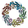
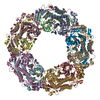
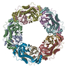
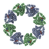
THIS IS A CHIMERICAL PROTEIN COMPOSED OF THE 29AA OF THE C-TERMINAL DOMAIN OF CAPA1 (RESIDUE NAME ...THIS IS A CHIMERICAL PROTEIN COMPOSED OF THE 29AA OF THE C-TERMINAL DOMAIN OF CAPA1 (RESIDUE NAME FROM 197 TO 222) IN FUSION WITH CAPB2 PROTEIN. THERE ARE TWO CHIMERICAL PROTEINS IN THE ASYMMETRIC UNIT
-
Experimental details
-
Experiment
Experiment
Method: X-RAY DIFFRACTION / Number of used crystals: 1
-
Sample preparation
Crystal
Density Matthews: 3.32 Å3/Da / Density % sol: 62.71 % / Description: NONE
Resolution: 2.6→19.79 Å / Cor.coef. Fo:Fc: 0.922 / Cor.coef. Fo:Fc free: 0.854 / SU B: 11.616 / SU ML: 0.241 / Cross valid method: THROUGHOUT / ESU R: 0.457 / ESU R Free: 0.307 / Stereochemistry target values: MAXIMUM LIKELIHOOD / Details: HYDROGENS HAVE BEEN ADDED IN THE RIDING POSITIONS
Rfactor
Num. reflection
% reflection
Selection details
Rfree
0.27
1164
5 %
RANDOM
Rwork
0.204
-
-
-
obs
0.208
22099
100 %
-
Solvent computation
Ion probe radii: 0.8 Å / Shrinkage radii: 0.8 Å / VDW probe radii: 1.4 Å / Solvent model: MASK
 Movie
Movie Controller


 Open data
 Basic information
 Components
 Keywords
 Function and homology information
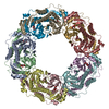
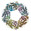
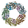
 STAPHYLOCOCCUS AUREUS (bacteria)
 X-RAY DIFFRACTION /
  Authors
 Citation
 Structure visualization
 Downloads & links
 Other downloads
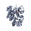


 PDBj
PDBj Assembly
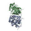

 Mass: 427.201 Da / Num. of mol.: 2 / Source method: obtained synthetically / Formula: C10H15N5O10P2 / Comment: ADP, energy-carrying molecule*YM
 Mass: 24.305 Da / Num. of mol.: 2 / Source method: obtained synthetically / Formula: Mg
 Mass: 18.015 Da / Num. of mol.: 112 / Source method: isolated from a natural source / Formula: H2O
 Sample preparation
 / Beamline: ID29 / Wavelength: 0.9
  Processing