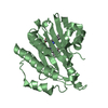
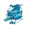
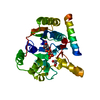
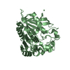
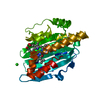
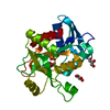
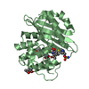
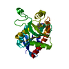
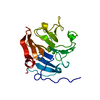
- PDB-3bfv: crystal structure of the chimerical protein CapAB -
+
Open data
ID or keywords:
Loading...
-
Basic information
Entry
Database: PDB / ID: 3bfv
Title
crystal structure of the chimerical protein CapAB
Components
Membrane protein CapA1, Protein tyrosine kinase,Protein tyrosine kinase
Keywords
TRANSFERASE / chimerical protein / P-loop protein / Capsule biogenesis/degradation / Exopolysaccharide synthesis / Membrane / Transmembrane / Virulence
Function / homology
Function and homology information
carbohydrate:proton symporter activity / polysaccharide transport / polysaccharide biosynthetic process / non-specific protein-tyrosine kinase / non-membrane spanning protein tyrosine kinase activity / protein tyrosine kinase activity / ATP binding / metal ion binding / identical protein binding / plasma membrane Similarity search - Function
A: Membrane protein CapA1, Protein tyrosine kinase,Protein tyrosine kinase B: Membrane protein CapA1, Protein tyrosine kinase,Protein tyrosine kinase hetero molecules
Resolution: 1.8→19.94 Å / Cor.coef. Fo:Fc: 0.942 / Cor.coef. Fo:Fc free: 0.914 / SU B: 2.532 / SU ML: 0.082 / Cross valid method: THROUGHOUT / ESU R: 0.142 / ESU R Free: 0.13 / Stereochemistry target values: MAXIMUM LIKELIHOOD / Details: HYDROGENS HAVE BEEN ADDED IN THE RIDING POSITIONS
Rfactor
Num. reflection
% reflection
Selection details
Rfree
0.217
2054
5 %
RANDOM
Rwork
0.178
-
-
-
obs
0.18
39022
100 %
-
Solvent computation
Ion probe radii: 0.8 Å / Shrinkage radii: 0.8 Å / VDW probe radii: 1.4 Å / Solvent model: MASK
Displacement parameters
Biso mean: 11.48 Å2
Baniso -1
Baniso -2
Baniso -3
1-
0 Å2
0 Å2
0 Å2
2-
-
0 Å2
0.01 Å2
3-
-
-
0.01 Å2
Refinement step
Cycle: LAST / Resolution: 1.8→19.94 Å
Protein
Nucleic acid
Ligand
Solvent
Total
Num. atoms
3728
0
56
418
4202
Refine LS restraints
Refine-ID
Type
Dev ideal
Dev ideal target
Number
X-RAY DIFFRACTION
r_bond_refined_d
0.008
0.022
3850
X-RAY DIFFRACTION
r_bond_other_d
X-RAY DIFFRACTION
r_angle_refined_deg
1.151
1.994
5242
X-RAY DIFFRACTION
r_angle_other_deg
X-RAY DIFFRACTION
r_dihedral_angle_1_deg
5.713
5
480
X-RAY DIFFRACTION
r_dihedral_angle_2_deg
35.772
25.802
162
X-RAY DIFFRACTION
r_dihedral_angle_3_deg
12.204
15
672
X-RAY DIFFRACTION
r_dihedral_angle_4_deg
20.082
15
16
X-RAY DIFFRACTION
r_chiral_restr
0.078
0.2
616
X-RAY DIFFRACTION
r_gen_planes_refined
0.003
0.02
2842
X-RAY DIFFRACTION
r_gen_planes_other
X-RAY DIFFRACTION
r_nbd_refined
0.186
0.2
1827
X-RAY DIFFRACTION
r_nbd_other
X-RAY DIFFRACTION
r_nbtor_refined
0.301
0.2
2731
X-RAY DIFFRACTION
r_nbtor_other
X-RAY DIFFRACTION
r_xyhbond_nbd_refined
0.094
0.2
363
X-RAY DIFFRACTION
r_xyhbond_nbd_other
X-RAY DIFFRACTION
r_metal_ion_refined
X-RAY DIFFRACTION
r_metal_ion_other
X-RAY DIFFRACTION
r_symmetry_vdw_refined
0.15
0.2
62
X-RAY DIFFRACTION
r_symmetry_vdw_other
X-RAY DIFFRACTION
r_symmetry_hbond_refined
0.122
0.2
48
X-RAY DIFFRACTION
r_symmetry_hbond_other
X-RAY DIFFRACTION
r_symmetry_metal_ion_refined
X-RAY DIFFRACTION
r_symmetry_metal_ion_other
X-RAY DIFFRACTION
r_mcbond_it
0.586
1.5
2484
X-RAY DIFFRACTION
r_mcbond_other
X-RAY DIFFRACTION
r_mcangle_it
0.935
2
3934
X-RAY DIFFRACTION
r_scbond_it
1.421
3
1532
X-RAY DIFFRACTION
r_scangle_it
2.231
4.5
1308
X-RAY DIFFRACTION
r_rigid_bond_restr
X-RAY DIFFRACTION
r_sphericity_free
X-RAY DIFFRACTION
r_sphericity_bonded
LS refinement shell
Resolution: 1.8→1.85 Å / Total num. of bins used: 20
Rfactor
Num. reflection
% reflection
Rfree
0.259
152
-
Rwork
0.224
2898
-
obs
-
-
100 %
+
About Yorodumi
-
News
-
Feb 9, 2022. New format data for meta-information of EMDB entries
New format data for meta-information of EMDB entries
Version 3 of the EMDB header file is now the official format.
The previous official version 1.9 will be removed from the archive.
In the structure databanks used in Yorodumi, some data are registered as the other names, "COVID-19 virus" and "2019-nCoV". Here are the details of the virus and the list of structure data.
Jan 31, 2019. EMDB accession codes are about to change! (news from PDBe EMDB page)
EMDB accession codes are about to change! (news from PDBe EMDB page)
The allocation of 4 digits for EMDB accession codes will soon come to an end. Whilst these codes will remain in use, new EMDB accession codes will include an additional digit and will expand incrementally as the available range of codes is exhausted. The current 4-digit format prefixed with “EMD-” (i.e. EMD-XXXX) will advance to a 5-digit format (i.e. EMD-XXXXX), and so on. It is currently estimated that the 4-digit codes will be depleted around Spring 2019, at which point the 5-digit format will come into force.
The EM Navigator/Yorodumi systems omit the EMD- prefix.
Related info.:Q: What is EMD? / ID/Accession-code notation in Yorodumi/EM Navigator
Yorodumi is a browser for structure data from EMDB, PDB, SASBDB, etc.
This page is also the successor to EM Navigator detail page, and also detail information page/front-end page for Omokage search.
The word "yorodu" (or yorozu) is an old Japanese word meaning "ten thousand". "mi" (miru) is to see.
Related info.:EMDB / PDB / SASBDB / Comparison of 3 databanks / Yorodumi Search / Aug 31, 2016. New EM Navigator & Yorodumi / Yorodumi Papers / Jmol/JSmol / Function and homology information / Changes in new EM Navigator and Yorodumi
 Movie
Movie Controller


 Open data
 Basic information
 Components
 Keywords
 Function and homology information
 Staphylococcus aureus (bacteria)
 X-RAY DIFFRACTION /
  Authors
 Citation
 Structure visualization
 Downloads & links
 Other downloads
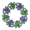


 PDBj
PDBj Assembly
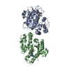


 Mass: 24.305 Da / Num. of mol.: 2 / Source method: obtained synthetically / Formula: Mg
 Mass: 427.201 Da / Num. of mol.: 2 / Source method: obtained synthetically / Formula: C10H15N5O10P2 / Comment: ADP, energy-carrying molecule*YM
 Mass: 18.015 Da / Num. of mol.: 418 / Source method: isolated from a natural source / Formula: H2O
 Sample preparation
 / Beamline: ID23-2 / Wavelength: 0.873
  Processing