 ムービー
ムービー コントローラー


+ データを開く

- 基本情報
基本情報
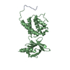
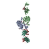
| 登録情報 | データベース: PDB / ID: 2vdk | ||||||||||||
|---|---|---|---|---|---|---|---|---|---|---|---|---|---|
| タイトル | Re-refinement of Integrin AlphaIIbBeta3 Headpiece | ||||||||||||
 要素 要素 |
| ||||||||||||
 キーワード キーワード | CELL ADHESION/IMMUNE SYSTEM / CELL ADHESION-IMMUNE SYSTEM COMPLEX / FIBRINOGEN BINDING / HOST-VIRUS INTERACTION / PYRROLIDONE CARBOXYLIC ACID / PHOSPHORYLATION | ||||||||||||
| 機能・相同性 |  機能・相同性情報 機能・相同性情報regulation of serotonin uptake / positive regulation of adenylate cyclase-inhibiting opioid receptor signaling pathway / tube development / alpha9-beta1 integrin-ADAM8 complex / regulation of trophoblast cell migration / integrin alphaIIb-beta3 complex / regulation of postsynaptic neurotransmitter receptor diffusion trapping / maintenance of postsynaptic specialization structure / alphav-beta3 integrin-vitronectin complex / regulation of extracellular matrix organization ...regulation of serotonin uptake / positive regulation of adenylate cyclase-inhibiting opioid receptor signaling pathway / tube development / alpha9-beta1 integrin-ADAM8 complex / regulation of trophoblast cell migration / integrin alphaIIb-beta3 complex / regulation of postsynaptic neurotransmitter receptor diffusion trapping / maintenance of postsynaptic specialization structure / alphav-beta3 integrin-vitronectin complex / regulation of extracellular matrix organization / platelet alpha granule membrane / positive regulation of glomerular mesangial cell proliferation / integrin alphav-beta3 complex / negative regulation of lipoprotein metabolic process / alphav-beta3 integrin-PKCalpha complex / fibrinogen binding / alphav-beta3 integrin-HMGB1 complex / negative regulation of lipid transport / vascular endothelial growth factor receptor 2 binding / positive regulation of vascular endothelial growth factor signaling pathway / Elastic fibre formation / cell-substrate junction assembly / alphav-beta3 integrin-IGF-1-IGF1R complex / positive regulation of bone resorption / platelet-derived growth factor receptor binding / mesodermal cell differentiation / angiogenesis involved in wound healing / filopodium membrane / regulation of release of sequestered calcium ion into cytosol / glycinergic synapse / extracellular matrix binding / positive regulation of vascular endothelial growth factor receptor signaling pathway / positive regulation of cell adhesion mediated by integrin / apolipoprotein A-I-mediated signaling pathway / regulation of bone resorption / negative regulation of low-density lipoprotein particle clearance / wound healing, spreading of epidermal cells / positive regulation of leukocyte migration / apoptotic cell clearance / positive regulation of fibroblast migration / integrin complex / cell adhesion mediated by integrin / Molecules associated with elastic fibres / smooth muscle cell migration / heterotypic cell-cell adhesion / positive regulation of smooth muscle cell migration / negative chemotaxis / Mechanical load activates signaling by PIEZO1 and integrins in osteocytes / positive regulation of cell-matrix adhesion / Syndecan interactions / p130Cas linkage to MAPK signaling for integrins / regulation of postsynaptic neurotransmitter receptor internalization / cellular response to insulin-like growth factor stimulus / positive regulation of osteoblast proliferation / protein disulfide isomerase activity / microvillus membrane / platelet-derived growth factor receptor signaling pathway / cell-substrate adhesion / PECAM1 interactions / GRB2:SOS provides linkage to MAPK signaling for Integrins / TGF-beta receptor signaling activates SMADs / lamellipodium membrane / fibronectin binding / negative regulation of macrophage derived foam cell differentiation / negative regulation of lipid storage / blood coagulation, fibrin clot formation / ECM proteoglycans / Integrin cell surface interactions / negative regulation of endothelial cell apoptotic process / positive regulation of T cell migration / coreceptor activity / cellular response to platelet-derived growth factor stimulus / Integrin signaling / positive regulation of endothelial cell proliferation / positive regulation of substrate adhesion-dependent cell spreading / substrate adhesion-dependent cell spreading / embryo implantation / positive regulation of endothelial cell migration / cell adhesion molecule binding / positive regulation of smooth muscle cell proliferation / Turbulent (oscillatory, disturbed) flow shear stress activates signaling by PIEZO1 and integrins in endothelial cells / cell-matrix adhesion / protein kinase C binding / response to activity / Signal transduction by L1 / integrin-mediated signaling pathway / regulation of actin cytoskeleton organization / wound healing / cellular response to mechanical stimulus / Signaling by high-kinase activity BRAF mutants / cell-cell adhesion / RUNX1 regulates genes involved in megakaryocyte differentiation and platelet function / platelet activation / MAP2K and MAPK activation / VEGFA-VEGFR2 Pathway / platelet aggregation / integrin binding / cellular response to xenobiotic stimulus / positive regulation of fibroblast proliferation / ruffle membrane 類似検索 - 分子機能 | ||||||||||||
| 生物種 |  HOMO SAPIENS (ヒト) HOMO SAPIENS (ヒト) | ||||||||||||
| 手法 |  X線回折 / シンクロトロン / 分子置換 / 解像度: 2.8 Å X線回折 / シンクロトロン / 分子置換 / 解像度: 2.8 Å | ||||||||||||
 データ登録者 データ登録者 | Springer, T.A. / Zhu, J. / Xiao, T. | ||||||||||||
 引用 引用 | ジャーナル: J.Cell Biol. / 年: 2008 タイトル: Structural Basis for Distinctive Recognition of Fibrinogen Gammac Peptide by the Platelet Integrin Alphaiibbeta3. 著者: Springer, T.A. / Zhu, J. / Xiao, T. #1: ジャーナル: Nature / 年: 2004タイトル: Structural Basis for Allostery in Integrins and Binding to Fibrinogen-Mimetic Therapeutics 著者: Xiao, T. / Takagi, J. / Coller, B.S. / Wang, J.-H. / Springer, T.A. | ||||||||||||
| 履歴 |
| ||||||||||||
| Remark 700 | SHEET THE SHEET STRUCTURE OF THIS MOLECULE IS BIFURCATED. IN ORDER TO REPRESENT THIS FEATURE IN ... SHEET THE SHEET STRUCTURE OF THIS MOLECULE IS BIFURCATED. IN ORDER TO REPRESENT THIS FEATURE IN THE SHEET RECORDS BELOW, TWO SHEETS ARE DEFINED. |
- 構造の表示
構造の表示
| 構造ビューア | 分子: MolmilJmol/JSmol |
|---|
- ダウンロードとリンク
-ダウンロード
| PDBx/mmCIF形式 | 2vdk.cif.gz | 297.8 KB | 表示 | PDBx/mmCIF形式 |
|---|---|---|---|---|
| PDB形式 | pdb2vdk.ent.gz | 237.6 KB | 表示 | PDB形式 |
| PDBx/mmJSON形式 | 2vdk.json.gz | ツリー表示 | PDBx/mmJSON形式 | |
| その他 |  その他のダウンロード その他のダウンロード |
-検証レポート
| アーカイブディレクトリ | https://data.pdbj.org/pub/pdb/validation_reports/vd/2vdkftp://data.pdbj.org/pub/pdb/validation_reports/vd/2vdk | HTTPS FTP |
|---|
-関連構造データ


-リンク
 PDBj
PDBj


- 集合体
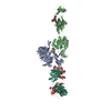
集合体
| 登録構造単位 | 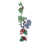
| ||||||||
|---|---|---|---|---|---|---|---|---|---|
| 1 |
| ||||||||
| 単位格子 |
|
-要素
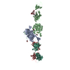
-タンパク質 , 2種, 2分子 AB
| #1: タンパク質 | 分子量: 49030.367 Da / 分子数: 1 / 断片: HEADPIECE, RESIDUES 32-483 / 由来タイプ: 組換発現 / 由来: (組換発現) HOMO SAPIENS (ヒト) / 細胞株 (発現宿主): LEC 3.2.8.1発現宿主:  CRICETULUS GRISEUS (モンゴルキヌゲネズミ) CRICETULUS GRISEUS (モンゴルキヌゲネズミ)参照: UniProt: P08514 |
|---|---|
| #2: タンパク質 | 分子量: 50969.664 Da / 分子数: 1 / 断片: HEADPIECE, RESIDUES 27-487 / 由来タイプ: 組換発現 / 由来: (組換発現) HOMO SAPIENS (ヒト) / 細胞株 (発現宿主): LEC 3.2.8.1発現宿主: CRICETULUS GRISEUS (モンゴルキヌゲネズミ)参照: UniProt: P05106 |
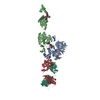
-抗体 , 2種, 2分子 HL
| #3: 抗体 | 分子量: 23766.473 Da / 分子数: 1 / 由来タイプ: 天然 / 由来: (天然) |
|---|---|
| #4: 抗体 | 分子量: 23332.686 Da / 分子数: 1 / 由来タイプ: 天然 / 由来: (天然) |
-糖 , 3種, 5分子 
| #5: 多糖 | alpha-D-mannopyranose-(1-3)-[alpha-D-mannopyranose-(1-6)]alpha-D-mannopyranose-(1-4)-2-acetamido-2- ...alpha-D-mannopyranose-(1-3)-[alpha-D-mannopyranose-(1-6)]alpha-D-mannopyranose-(1-4)-2-acetamido-2-deoxy-beta-D-glucopyranose-(1-4)-2-acetamido-2-deoxy-beta-D-glucopyranose |
|---|---|
| #6: 多糖 | alpha-D-mannopyranose-(1-3)-[alpha-D-mannopyranose-(1-6)]alpha-D-mannopyranose-(1-6)-[alpha-D- ...alpha-D-mannopyranose-(1-3)-[alpha-D-mannopyranose-(1-6)]alpha-D-mannopyranose-(1-6)-[alpha-D-mannopyranose-(1-3)]alpha-D-mannopyranose-(1-4)-2-acetamido-2-deoxy-beta-D-glucopyranose-(1-4)-2-acetamido-2-deoxy-beta-D-glucopyranose |
| #9: 糖 |  タイプ: D-saccharide, beta linking / 分子量: 221.208 Da / 分子数: 3 / 由来タイプ: 組換発現 / 式: C8H15NO6 タイプ: D-saccharide, beta linking / 分子量: 221.208 Da / 分子数: 3 / 由来タイプ: 組換発現 / 式: C8H15NO6 |
-非ポリマー , 5種, 622分子 


| #7: 化合物 |  分子量: 92.094 Da / 分子数: 2 / 由来タイプ: 合成 / 式: C3H8O3 分子量: 92.094 Da / 分子数: 2 / 由来タイプ: 合成 / 式: C3H8O3#8: 化合物 | ChemComp-CA /  分子量: 40.078 Da / 分子数: 6 / 由来タイプ: 合成 / 式: Ca 分子量: 40.078 Da / 分子数: 6 / 由来タイプ: 合成 / 式: Ca#10: 化合物 | ChemComp-CAC / |  分子量: 136.989 Da / 分子数: 1 / 由来タイプ: 合成 / 式: C2H6AsO2 分子量: 136.989 Da / 分子数: 1 / 由来タイプ: 合成 / 式: C2H6AsO2#11: 化合物 | ChemComp-MG / |  分子量: 24.305 Da / 分子数: 1 / 由来タイプ: 合成 / 式: Mg 分子量: 24.305 Da / 分子数: 1 / 由来タイプ: 合成 / 式: Mg#12: 水 | ChemComp-HOH / | 分子量: 18.015 Da / 分子数: 612 / 由来タイプ: 天然 / 式: H2O |
|---|
-詳細
| Has protein modification | Y |
|---|---|
| 非ポリマーの詳細 | CACODYLATE ION (CAC): CACODYLATE IS FOUND IN THE LIGAND BINDING SITE. BASED ON IMPURITIES IN ...CACODYLATE |
| 配列の詳細 | ACCORDING TO THE AUTHORS THE CORRECT CHAIN A SEQUENCE IS ANNOTATED IN NCBI ENTRY GI 88758615 WHICH ...ACCORDING TO THE AUTHORS THE CORRECT CHAIN A SEQUENCE IS ANNOTATED IN NCBI ENTRY GI 88758615 WHICH SHOULD BE USED IN PLACE OF P08514. |
-実験情報
-実験
| 実験 | 手法: X線回折 / 使用した結晶の数: 1 |
|---|
- 試料調製
試料調製
| 結晶 | マシュー密度: 3.7 Å3/Da / 溶媒含有率: 66.8 % / 解説: NONE |
|---|---|
| 結晶化 | 温度: 277 K / 手法: 蒸気拡散法, ハンギングドロップ法 / pH: 6.5 詳細: 11% PEG 3350, 0.7 M MAGNESIUM ACETATE, 0.1 M SODIUM CACODYLATE, PH 6.5, VAPOR DIFFUSION, HANGING DROP, TEMPERATURE 277K |
-データ収集
| 回折 | 平均測定温度: 100 K |
|---|---|
| 放射光源 | 由来: シンクロトロン / サイト: APS  / ビームライン: 19-ID / 波長: 0.9793 / ビームライン: 19-ID / 波長: 0.9793 |
| 検出器 | タイプ: CUSTOM (SBC2 3K) / 検出器: CCD / 日付: 2003年8月5日 / 詳細: MIRROR |
| 放射 | モノクロメーター: SI(111) / プロトコル: SINGLE WAVELENGTH / 単色(M)・ラウエ(L): M / 散乱光タイプ: x-ray |
| 放射波長 | 波長: 0.9793 Å / 相対比: 1 |
| 反射 | 解像度: 2.8→50 Å / Num. obs: 55875 / % possible obs: 99.9 % / Observed criterion σ(I): 0 / 冗長度: 7.5 % / Rmerge(I) obs: 0.13 / Net I/σ(I): 14 |
| 反射 シェル | 解像度: 2.8→2.9 Å / Rmerge(I) obs: 0.68 / Mean I/σ(I) obs: 3.1 / % possible all: 99.9 |
- 解析
解析
| ソフトウェア |
| ||||||||||||||||||||||||||||||||||||||||||||||||||||||||||||||||||||||||||||||||||||||||||||||||||||||||||||||||||||||||||||||||||||||||||||||||||||||||||||||||||||||||||||||||||||||
|---|---|---|---|---|---|---|---|---|---|---|---|---|---|---|---|---|---|---|---|---|---|---|---|---|---|---|---|---|---|---|---|---|---|---|---|---|---|---|---|---|---|---|---|---|---|---|---|---|---|---|---|---|---|---|---|---|---|---|---|---|---|---|---|---|---|---|---|---|---|---|---|---|---|---|---|---|---|---|---|---|---|---|---|---|---|---|---|---|---|---|---|---|---|---|---|---|---|---|---|---|---|---|---|---|---|---|---|---|---|---|---|---|---|---|---|---|---|---|---|---|---|---|---|---|---|---|---|---|---|---|---|---|---|---|---|---|---|---|---|---|---|---|---|---|---|---|---|---|---|---|---|---|---|---|---|---|---|---|---|---|---|---|---|---|---|---|---|---|---|---|---|---|---|---|---|---|---|---|---|---|---|---|---|
| 精密化 | 構造決定の手法: 分子置換 開始モデル: PDB ENTRY 1TXV  1txv 解像度: 2.8→42.64 Å / Cor.coef. Fo:Fc: 0.951 / Cor.coef. Fo:Fc free: 0.924 / SU B: 17.783 / SU ML: 0.187 / TLS residual ADP flag: UNVERIFIED / 交差検証法: THROUGHOUT / ESU R: 0.648 / ESU R Free: 0.279 / 立体化学のターゲット値: MAXIMUM LIKELIHOOD 詳細: HYDROGENS HAVE BEEN ADDED IN THE RIDING POSITIONS. THESE ARE RE-REFINED COORDINATES OF THE PREVIOUS WWPDB SUBMISSION 1TY3. THE STARTING MODEL WAS A 2.4 ANGSTROM STRUCTURE WITH A DIFFERENT ...詳細: HYDROGENS HAVE BEEN ADDED IN THE RIDING POSITIONS. THESE ARE RE-REFINED COORDINATES OF THE PREVIOUS WWPDB SUBMISSION 1TY3. THE STARTING MODEL WAS A 2.4 ANGSTROM STRUCTURE WITH A DIFFERENT BOUND LIGAND. THE MODEL IS REFINED TO LOWER RFREE. ONE SEQUENCE MISTAKE IN THE AIIB SUBUNIT IS CORRECTED. MORE OF BETA SUBUNIT DOMAIN I-EGF1 IS BUILT. MISTAKES IN CARBOHYDRATE ANOMERIC LINKAGES ARE ALSO CORRECTED.
| ||||||||||||||||||||||||||||||||||||||||||||||||||||||||||||||||||||||||||||||||||||||||||||||||||||||||||||||||||||||||||||||||||||||||||||||||||||||||||||||||||||||||||||||||||||||
| 溶媒の処理 | イオンプローブ半径: 0.8 Å / 減衰半径: 0.8 Å / VDWプローブ半径: 1.2 Å / 溶媒モデル: BABINET MODEL WITH MASK | ||||||||||||||||||||||||||||||||||||||||||||||||||||||||||||||||||||||||||||||||||||||||||||||||||||||||||||||||||||||||||||||||||||||||||||||||||||||||||||||||||||||||||||||||||||||
| 原子変位パラメータ | Biso mean: 17.09 Å2
| ||||||||||||||||||||||||||||||||||||||||||||||||||||||||||||||||||||||||||||||||||||||||||||||||||||||||||||||||||||||||||||||||||||||||||||||||||||||||||||||||||||||||||||||||||||||
| 精密化ステップ | サイクル: LAST / 解像度: 2.8→42.64 Å
| ||||||||||||||||||||||||||||||||||||||||||||||||||||||||||||||||||||||||||||||||||||||||||||||||||||||||||||||||||||||||||||||||||||||||||||||||||||||||||||||||||||||||||||||||||||||
| 拘束条件 |
|