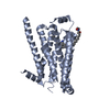
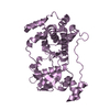
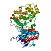
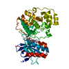
SHEET THE SHEET STRUCTURE OF THIS MOLECULE IS BIFURCATED. IN ORDER TO REPRESENT THIS FEATURE IN ... SHEET THE SHEET STRUCTURE OF THIS MOLECULE IS BIFURCATED. IN ORDER TO REPRESENT THIS FEATURE IN THE SHEET RECORDS BELOW, TWO SHEETS ARE DEFINED.
Mass: 18.015 Da / Num. of mol.: 174 / Source method: isolated from a natural source / Formula: H2O
Compound details
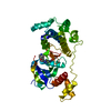
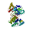
ENGINEERED RESIDUE IN CHAIN A, THR 163 TO ASN ENGINEERED RESIDUE IN CHAIN A, ARG 164 TO GLN
Sequence details
AMINO ACIDS 163 AND 164 TR REPLACED BY NQ
-
Experimental details
-
Experiment
Experiment
Method: X-RAY DIFFRACTION / Number of used crystals: 1
-
Sample preparation
Crystal
Density Matthews: 2.37 Å3/Da / Density % sol: 48 % / Description: NONE
Crystal grow
pH: 8.5 Details: 1 MICROLITER OF 15 MG/ML RECOMBINANT RAT CBG AND 1 MICROLITER OF RESERVOIR SOLUTION (30% (W/V) PEG 4000, 300 MM LI2SO4, 100 MM TRIS-HCL, PH 8.5) AGAINST 700 MICROLITER OF RESERVOIR SOLUTION
Resolution: 1.93→37.42 Å / Cor.coef. Fo:Fc: 0.954 / Cor.coef. Fo:Fc free: 0.923 / SU B: 10.9 / SU ML: 0.154 / Cross valid method: THROUGHOUT / ESU R: 0.18 / ESU R Free: 0.171 / Stereochemistry target values: MAXIMUM LIKELIHOOD Details: HYDROGENS HAVE BEEN ADDED IN THE RIDING POSITIONS. ZERO OCCUPANCIES SET FOR SIDE CHAINS WHERE NO DENSITY WAS SEEN.
Rfactor
Num. reflection
% reflection
Selection details
Rfree
0.264
2328
8 %
RANDOM
Rwork
0.21
-
-
-
obs
0.214
26770
100 %
-
Solvent computation
Ion probe radii: 0.8 Å / Shrinkage radii: 0.8 Å / VDW probe radii: 1.2 Å / Solvent model: BABINET MODEL WITH MASK
 Movie
Movie Controller

 Yorodumi
 Open data
 Basic information
 Components
 Keywords
 Function and homology information
 X-RAY DIFFRACTION /
  Authors
 Citation
 Structure visualization
 Downloads & links
 Other downloads

 PDBj
PDBj

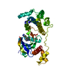
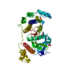
 Assembly
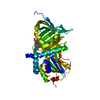


 Mass: 362.460 Da / Num. of mol.: 1 / Source method: obtained synthetically / Formula: C21H30O5 / Comment: medication, hormone*YM
 Mass: 18.015 Da / Num. of mol.: 174 / Source method: isolated from a natural source / Formula: H2O
 Sample preparation
 / Beamline: 14.1 / Wavelength: 0.9537
  Processing