 Movie
Movie Controller

[English] 日本語
 Yorodumi
- PDB-2v8f: Mouse Profilin IIa in complex with a double repeat from the FH1 d... -
+ Open data

- Basic information
| Entry | Database: PDB / ID: 2v8f | ||||||
|---|---|---|---|---|---|---|---|
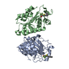
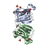
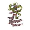
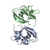
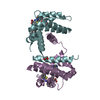
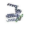
| Title | Mouse Profilin IIa in complex with a double repeat from the FH1 domain of mDia1 | ||||||
 Components Components |
| ||||||
 Keywords Keywords | PROTEIN BINDING / ALTERNATIVE SPLICING / PROTEIN-BINDING / CYTOPLASM / ACETYLATION / CYTOSKELETON / ACTIN-BINDING | ||||||
| Function / homology |  Function and homology information Function and homology informationSignaling by ROBO receptors / negative regulation of neuron projection regeneration / multicellular organismal locomotion / presynaptic actin cytoskeleton organization / negative regulation of ruffle assembly / ERBB2 Regulates Cell Motility / RHOF GTPase cycle / RHOC GTPase cycle / : / neuron projection retraction ...Signaling by ROBO receptors / negative regulation of neuron projection regeneration / multicellular organismal locomotion / presynaptic actin cytoskeleton organization / negative regulation of ruffle assembly / ERBB2 Regulates Cell Motility / RHOF GTPase cycle / RHOC GTPase cycle / : / neuron projection retraction / actin nucleation / RHOB GTPase cycle / negative regulation of epithelial cell migration / RHOA GTPase cycle / protein localization to microtubule / negative regulation of actin filament polymerization / modification of postsynaptic actin cytoskeleton / RHO GTPases Activate Formins / profilin binding / positive regulation of actin filament bundle assembly / regulation of actin filament polymerization / cellular response to histamine / regulation of release of sequestered calcium ion into cytosol / regulation of microtubule-based process / axon midline choice point recognition / regulation of cytoskeleton organization / regulation of synaptic vesicle exocytosis / positive regulation of actin filament polymerization / brush border / actin monomer binding / ephrin receptor signaling pathway / synaptic vesicle endocytosis / positive regulation of stress fiber assembly / actin filament polymerization / Neutrophil degranulation / phosphatidylinositol-4,5-bisphosphate binding / cytoskeleton organization / presynaptic modulation of chemical synaptic transmission / actin filament / sensory perception of sound / brain development / modulation of chemical synaptic transmission / Schaffer collateral - CA1 synapse / small GTPase binding / ruffle membrane / neuron projection development / terminal bouton / mitotic spindle / intracellular protein localization / regulation of cell shape / presynapse / actin binding / actin cytoskeleton organization / gene expression / cytoskeleton / transmembrane transporter binding / neuron projection / protein stabilization / postsynapse / positive regulation of cell migration / centrosome / glutamatergic synapse / ATP hydrolysis activity / identical protein binding / nucleus / cytoplasm / cytosol Similarity search - Function | ||||||
| Biological species |  | ||||||
| Method |  X-RAY DIFFRACTION / SYNCHROTRON / MOLECULAR REPLACEMENT / Resolution: 1.1 Å X-RAY DIFFRACTION / SYNCHROTRON / MOLECULAR REPLACEMENT / Resolution: 1.1 Å | ||||||
 Authors Authors | Kursula, P. / Kursula, I. / Downer, J. / Witke, W. / Wilmanns, M. | ||||||
 Citation Citation | Journal: J.Mol.Biol. / Year: 2008 Title: High-Resolution Structural Analysis of Mammalian Profilin 2A Complex Formation with Two Physiological Ligands: The Formin Homology 1 Domain of Mdia1 and the Proline-Rich Domain of Vasp. Authors: Kursula, P. / Kursula, I. / Massimi, M. / Song, Y.H. / Downer, J. / Stanley, W.A. / Witke, W. / Wilmanns, M. | ||||||
| History |
|
- Structure visualization
| Structure viewer | Molecule: MolmilJmol/JSmol |
|---|
- Downloads & links
-Download
| PDBx/mmCIF format | 2v8f.cif.gz | 145.7 KB | Display | PDBx/mmCIF format |
|---|---|---|---|---|
| PDB format | pdb2v8f.ent.gz | 115.2 KB | Display | PDB format |
| PDBx/mmJSON format | 2v8f.json.gz | Tree view | PDBx/mmJSON format | |
| Others |  Other downloads Other downloads |
-Validation report
| Arichive directory | https://data.pdbj.org/pub/pdb/validation_reports/v8/2v8fftp://data.pdbj.org/pub/pdb/validation_reports/v8/2v8f | HTTPS FTP |
|---|
-Related structure data
| Related structure data | 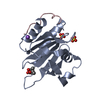 2v8cC  1d1jS C: citing same article ( S: Starting model for refinement |
|---|---|
| Similar structure data |


-Links
 PDBj
PDBj


- Assembly
| Deposited unit | 
| ||||||||
|---|---|---|---|---|---|---|---|---|---|
| 1 |
| ||||||||
| Unit cell |
|
-Components
-Protein / Protein/peptide , 2 types, 3 molecules ABC
| #1: Protein | Mass: 15064.216 Da / Num. of mol.: 2 Source method: isolated from a genetically manipulated source Source: (gene. exp.)  #2: Protein/peptide | | Mass: 2007.414 Da / Num. of mol.: 1 / Fragment: FH1 DOMAIN, RESIDUES 635-655 / Source method: obtained synthetically Details: TWO PROLINE-RICH REPEATS DERIVED FROM THE MOUSE HOMOLOGUE OF DIAPHANOUS 1 (MDIA1). Source: (synth.) |
|---|
-Non-polymers , 5 types, 365 molecules 


| #3: Chemical | ChemComp-SO4 /  Mass: 96.063 Da / Num. of mol.: 5 / Source method: obtained synthetically / Formula: SO4 Mass: 96.063 Da / Num. of mol.: 5 / Source method: obtained synthetically / Formula: SO4#4: Chemical | ChemComp-NA / |  Mass: 22.990 Da / Num. of mol.: 1 / Source method: obtained synthetically / Formula: Na Mass: 22.990 Da / Num. of mol.: 1 / Source method: obtained synthetically / Formula: Na#5: Chemical | ChemComp-GOL / |  Mass: 92.094 Da / Num. of mol.: 1 / Source method: obtained synthetically / Formula: C3H8O3 Mass: 92.094 Da / Num. of mol.: 1 / Source method: obtained synthetically / Formula: C3H8O3#6: Chemical | ChemComp-IPA / |  Mass: 60.095 Da / Num. of mol.: 1 / Source method: obtained synthetically / Formula: C3H8O Mass: 60.095 Da / Num. of mol.: 1 / Source method: obtained synthetically / Formula: C3H8O#7: Water | ChemComp-HOH / | Mass: 18.015 Da / Num. of mol.: 357 / Source method: isolated from a natural source / Formula: H2O |
|---|
-Details
| Has protein modification | Y |
|---|
-Experimental details
-Experiment
| Experiment | Method: X-RAY DIFFRACTION |
|---|
- Sample preparation
| Crystal | Density Matthews: 1.65 Å3/Da / Density % sol: 25.01 % / Description: NONE |
|---|
-Data collection
| Diffraction | Mean temperature: 100 K |
|---|---|
| Diffraction source | Source: SYNCHROTRON / Site: BESSY  / Beamline: 14.1 / Wavelength: 1.0408 / Beamline: 14.1 / Wavelength: 1.0408 |
| Detector | Type: MARRESEARCH / Detector: CCD |
| Radiation | Protocol: SINGLE WAVELENGTH / Monochromatic (M) / Laue (L): M / Scattering type: x-ray |
| Radiation wavelength | Wavelength: 1.0408 Å / Relative weight: 1 |
| Reflection | Resolution: 1.1→20 Å / Num. obs: 93713 / % possible obs: 92.8 % / Observed criterion σ(I): -3 / Redundancy: 3.2 % / Rmerge(I) obs: 0.08 / Net I/σ(I): 9.3 |
| Reflection shell | Resolution: 1.1→1.2 Å / Redundancy: 2.5 % / Rmerge(I) obs: 0.4 / Mean I/σ(I) obs: 2.9 / % possible all: 75.4 |
- Processing
| Software |
| ||||||||||||||||||||
|---|---|---|---|---|---|---|---|---|---|---|---|---|---|---|---|---|---|---|---|---|---|
| Refinement | Method to determine structure: MOLECULAR REPLACEMENT Starting model: PDB ENTRY 1D1J Resolution: 1.1→20 Å / Cross valid method: THROUGHOUT / σ(F): 0 Details: FINAL ROUND OF B FACTOR REFINEMENT DONE IN PHENIX.REFINE
| ||||||||||||||||||||
| Refinement step | Cycle: LAST / Resolution: 1.1→20 Å
|
