 Movie
Movie Controller

[English] 日本語
 Yorodumi
- PDB-2v7w: X-ray crystal structure of 5'-fluorodeoxyadenosine synthase s158g... -
+ Open data

- Basic information
| Entry | Database: PDB / ID: 2v7w | |||||||||
|---|---|---|---|---|---|---|---|---|---|---|
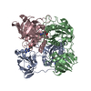
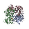
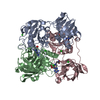
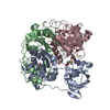
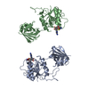
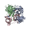
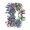
| Title | X-ray crystal structure of 5'-fluorodeoxyadenosine synthase s158g mutant complexed with 5'-fluorodeoxyadenosin | |||||||||
 Components Components | 5'-FLUORO-5'-DEOXYADENOSINE SYNTHASE | |||||||||
 Keywords Keywords | TRANSFERASE / FLUORINATION / BIOSYNTHETIC PROTEIN | |||||||||
| Function / homology |  Function and homology information Function and homology information | |||||||||
| Biological species |  STREPTOMYCES CATTLEYA (bacteria) STREPTOMYCES CATTLEYA (bacteria) | |||||||||
| Method |  X-RAY DIFFRACTION / SYNCHROTRON / MOLECULAR REPLACEMENT / Resolution: 1.9 Å X-RAY DIFFRACTION / SYNCHROTRON / MOLECULAR REPLACEMENT / Resolution: 1.9 Å | |||||||||
 Authors Authors | Zhu, X. / O'Hagan, D. / Naismith, J.H. | |||||||||
 Citation Citation | Journal: J. Am. Chem. Soc. / Year: 2007 Title: Mechanism of enzymatic fluorination in Streptomyces cattleya. Authors: Zhu, X. / Robinson, D.A. / McEwan, A.R. / O'Hagan, D. / Naismith, J.H. | |||||||||
| History |
| |||||||||
| Remark 700 | SHEET DETERMINATION METHOD: DSSP THE SHEETS PRESENTED AS "AB", "BB" IN EACH CHAIN ON SHEET RECORDS ... SHEET DETERMINATION METHOD: DSSP THE SHEETS PRESENTED AS "AB", "BB" IN EACH CHAIN ON SHEET RECORDS BELOW IS ACTUALLY AN 8-STRANDED BARREL THIS IS REPRESENTED BY A 9-STRANDED SHEET IN WHICH THE FIRST AND LAST STRANDS ARE IDENTICAL. THE SHEETS PRESENTED AS "CB" IN EACH CHAIN ON SHEET RECORDS BELOW IS ACTUALLY AN 7-STRANDED BARREL THIS IS REPRESENTED BY A 8-STRANDED SHEET IN WHICH THE FIRST AND LAST STRANDS ARE IDENTICAL. |
- Structure visualization
| Structure viewer | Molecule: MolmilJmol/JSmol |
|---|
- Downloads & links
-Download
| PDBx/mmCIF format | 2v7w.cif.gz | 187.3 KB | Display | PDBx/mmCIF format |
|---|---|---|---|---|
| PDB format | pdb2v7w.ent.gz | 150.8 KB | Display | PDB format |
| PDBx/mmJSON format | 2v7w.json.gz | Tree view | PDBx/mmJSON format | |
| Others |  Other downloads Other downloads |
-Validation report
| Summary document | 2v7w_validation.pdf.gz | 1.3 MB | Display | wwPDB validaton report |
|---|---|---|---|---|
| Full document | 2v7w_full_validation.pdf.gz | 1.3 MB | Display | |
| Data in XML | 2v7w_validation.xml.gz | 38.5 KB | Display | |
| Data in CIF | 2v7w_validation.cif.gz | 55.7 KB | Display | |
| Arichive directory | https://data.pdbj.org/pub/pdb/validation_reports/v7/2v7wftp://data.pdbj.org/pub/pdb/validation_reports/v7/2v7w | HTTPS FTP |
-Related structure data
| Related structure data | 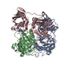 2v7tC 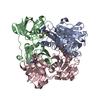 2v7uC 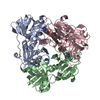 2v7vC 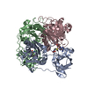 2v7xC  1rqpS S: Starting model for refinement C: citing same article ( |
|---|---|
| Similar structure data |


-Links
 PDBj
PDBj- Assembly
| Deposited unit | 
| ||||||||||||||||||||||||||||||||||||||||||||||||||||||||||||||||||||||||||||||||||||||||||||||||||||||||||||||||||||||||||||||||||||||||||||||||||||||
|---|---|---|---|---|---|---|---|---|---|---|---|---|---|---|---|---|---|---|---|---|---|---|---|---|---|---|---|---|---|---|---|---|---|---|---|---|---|---|---|---|---|---|---|---|---|---|---|---|---|---|---|---|---|---|---|---|---|---|---|---|---|---|---|---|---|---|---|---|---|---|---|---|---|---|---|---|---|---|---|---|---|---|---|---|---|---|---|---|---|---|---|---|---|---|---|---|---|---|---|---|---|---|---|---|---|---|---|---|---|---|---|---|---|---|---|---|---|---|---|---|---|---|---|---|---|---|---|---|---|---|---|---|---|---|---|---|---|---|---|---|---|---|---|---|---|---|---|---|---|---|---|
| 1 | 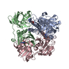
| ||||||||||||||||||||||||||||||||||||||||||||||||||||||||||||||||||||||||||||||||||||||||||||||||||||||||||||||||||||||||||||||||||||||||||||||||||||||
| Unit cell |
| ||||||||||||||||||||||||||||||||||||||||||||||||||||||||||||||||||||||||||||||||||||||||||||||||||||||||||||||||||||||||||||||||||||||||||||||||||||||
| Noncrystallographic symmetry (NCS) | NCS domain:
NCS domain segments: Ens-ID: 1 / Refine code: 2
|