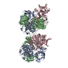
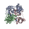
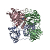
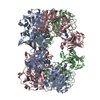
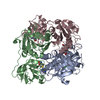
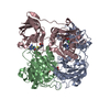
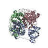
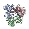
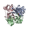
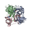
Entry Database : PDB / ID : 2c2wTitle The fluorinase from Streptomyces cattleya is also a chlorinase. Structure of 5'-chloro-5'-deoxyadenosine crystallised in the fluorinase. 5'-FLUORO-5'-DEOXYADENOSINE SYNTHASE Keywords / / / / / / / Function / homology Function Domain/homology Component
/ / / / / / / / / / / / / / / / / / / / / / / / Biological species STREPTOMYCES CATTLEYA (bacteria)Method / / / Resolution : 2 Å Authors McEwan, A.R. / Naismith, J.H. / O'Hagan, D. Journal : Angew. Chem. Int. Ed. Engl. / Year : 2006Title : The fluorinase from Streptomyces cattleya is also a chlorinase.Authors : Deng, H. / Cobb, S.L. / McEwan, A.R. / McGlinchey, R.P. / Naismith, J.H. / O'Hagan, D. / Robinson, D.A. / Spencer, J.B. History Deposition Sep 30, 2005 Deposition site / Processing site Revision 1.0 Dec 19, 2005 Provider / Type Revision 1.1 Jul 13, 2011 Group / Version format complianceRevision 1.2 Mar 28, 2018 Group / Structure summary / Category / citation / citation_authorItem _audit_author.name / _citation.journal_abbrev ... _audit_author.name / _citation.journal_abbrev / _citation.page_last / _citation.pdbx_database_id_DOI / _citation.title / _citation_author.name Revision 1.3 Dec 13, 2023 Group Data collection / Database references ... Data collection / Database references / Derived calculations / Other / Refinement description Category chem_comp_atom / chem_comp_bond ... chem_comp_atom / chem_comp_bond / database_2 / pdbx_database_status / pdbx_initial_refinement_model / struct_site Item _database_2.pdbx_DOI / _database_2.pdbx_database_accession ... _database_2.pdbx_DOI / _database_2.pdbx_database_accession / _pdbx_database_status.status_code_sf / _struct_site.pdbx_auth_asym_id / _struct_site.pdbx_auth_comp_id / _struct_site.pdbx_auth_seq_id
Show all Show less Remark 700 SHEET DETERMINATION METHOD: DSSP THE SHEETS PRESENTED AS "AB" IN EACH CHAIN ON SHEET RECORDS BELOW ... SHEET DETERMINATION METHOD: DSSP THE SHEETS PRESENTED AS "AB" IN EACH CHAIN ON SHEET RECORDS BELOW IS ACTUALLY AN 8-STRANDED BARREL THIS IS REPRESENTED BY A 9-STRANDED SHEET IN WHICH THE FIRST AND LAST STRANDS ARE IDENTICAL. THE SHEETS PRESENTED AS "BB" IN EACH CHAIN ON SHEET RECORDS BELOW IS ACTUALLY AN 7-STRANDED BARREL THIS IS REPRESENTED BY A 8-STRANDED SHEET IN WHICH THE FIRST AND LAST STRANDS ARE IDENTICAL. THE SHEETS PRESENTED AS "CB" IN EACH CHAIN ON SHEET RECORDS BELOW IS ACTUALLY AN 8-STRANDED BARREL THIS IS REPRESENTED BY A 9-STRANDED SHEET IN WHICH THE FIRST AND LAST STRANDS ARE IDENTICAL.
 Movie
Movie Controller

 Yorodumi
 Open data
 Basic information
 Components
 Keywords
 Function and homology information
 STREPTOMYCES CATTLEYA (bacteria)
 X-RAY DIFFRACTION /
  Authors
 Citation
 Structure visualization
 Downloads & links
 Other downloads
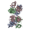


 PDBj
PDBj Assembly

 Mass: 285.687 Da / Num. of mol.: 3 / Source method: obtained synthetically / Formula: C10H12ClN5O3
 Mass: 35.453 Da / Num. of mol.: 7 / Source method: obtained synthetically / Formula: Cl
 Mass: 18.015 Da / Num. of mol.: 559 / Source method: isolated from a natural source / Formula: H2O
 Sample preparation
 / Beamline: ID14-2 / Wavelength: 0.933
  Processing