[English] 日本語
Yorodumi
- PDB-2v66: Crystal Structure of the coiled-coil domain of Ndel1 (a.a. 58 to ... -

+
Open data


ID or keywords:

Loading...

-
Basic information

Entry
Database: PDB / ID: 2v66
Title
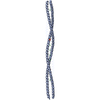
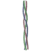
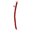
Crystal Structure of the coiled-coil domain of Ndel1 (a.a. 58 to 169) C
ComponentsNUCLEAR DISTRIBUTION PROTEIN NUDE-LIKE 1
KeywordsSTRUCTURAL PROTEIN / DEVELOPMENTAL PROTEIN / STRUCTURAL PROTEIN PHOSPHORYLATION / TRANSPORT / MICROTUBULE / NEUROGENESIS / CYTOSKELETON / DIFFERENTIATION
Function / homology
Function and homology information


: / neurofilament cytoskeleton / cerebral cortex radially oriented cell migration / kinetochore => GO:0000776 / establishment of chromosome localization / central nervous system neuron axonogenesis / central region of growth cone / oligopeptidase activity / nuclear membrane disassembly / vesicle transport along microtubule ...: / neurofilament cytoskeleton / cerebral cortex radially oriented cell migration / kinetochore => GO:0000776 / establishment of chromosome localization / central nervous system neuron axonogenesis / central region of growth cone / oligopeptidase activity / nuclear membrane disassembly / vesicle transport along microtubule / neurofilament cytoskeleton organization / lysosome localization / microtubule nucleation / axon hillock / retrograde axonal transport / activation of GTPase activity / positive regulation of ruffle assembly / mitotic centrosome separation / centrosome localization / inner cell mass cell proliferation / neuron projection extension / kinesin complex / positive regulation of axon regeneration / regulation of intracellular protein transport / regulation of neuron projection development / beta-tubulin binding / cell leading edge / establishment of mitotic spindle orientation / alpha-tubulin binding / positive regulation of axon extension / Amplification of signal from unattached kinetochores via a MAD2 inhibitory signal / Mitotic Prometaphase / EML4 and NUDC in mitotic spindle formation / axon cytoplasm / Resolution of Sister Chromatid Cohesion / positive regulation of GTPase activity / chromosome segregation / RHO GTPases Activate Formins / neuron migration / kinetochore / spindle / Separation of Sister Chromatids / cell migration / synaptic vesicle / insulin receptor signaling pathway / nuclear envelope / microtubule binding / microtubule / centrosome / protein-containing complex binding / identical protein binding / cytosol
Similarity search - Function
Single alpha-helices involved in coiled-coils or other helix-helix interfaces - #1080 / Nuclear distribution protein nudE-like 1 / NUDE domain / NUDE family / NUDE protein, C-terminal conserved region / Single alpha-helices involved in coiled-coils or other helix-helix interfaces / Helix non-globular / Special
Similarity search - Domain/homology
: / Nuclear distribution protein nudE-like 1
Similarity search - Component
Biological speciesHOMO SAPIENS (human)
MethodX-RAY DIFFRACTION / SYNCHROTRON / MAD / Resolution: 2.1 Å
AuthorsTarricone, C. / Perrina, F. / Musacchio, A.
CitationJournal: Structure / Year: 2007
Title: The Structure of the Coiled-Coil Domain of Ndel1 and the Basis of its Interaction with Lis1, the Causal Protein of Miller-Dieker Lissencephaly.
Authors: Derewenda, U. / Tarricone, C. / Choi, W.C. / Cooper, D.R. / Lukasik, S. / Perrina, F. / Tripathy, A. / Kim, M.H. / Cafiso, D.S. / Musacchio, A. / Derewenda, Z.S.
History
DepositionJul 13, 2007Deposition site: PDBE / Processing site: PDBE
Revision 1.0Nov 27, 2007Provider: repository / Type: Initial release
Revision 1.1Jul 13, 2011Group: Advisory / Refinement description / Version format compliance
Revision 1.2May 8, 2024Group: Data collection / Database references ...Data collection / Database references / Derived calculations / Other
Category: chem_comp_atom / chem_comp_bond ...chem_comp_atom / chem_comp_bond / database_2 / pdbx_database_status / struct_conn
Item: _database_2.pdbx_DOI / _database_2.pdbx_database_accession ..._database_2.pdbx_DOI / _database_2.pdbx_database_accession / _pdbx_database_status.status_code_sf / _struct_conn.ptnr1_auth_comp_id / _struct_conn.ptnr1_auth_seq_id / _struct_conn.ptnr1_label_asym_id / _struct_conn.ptnr1_label_atom_id / _struct_conn.ptnr1_label_comp_id / _struct_conn.ptnr1_label_seq_id / _struct_conn.ptnr1_symmetry / _struct_conn.ptnr2_auth_comp_id / _struct_conn.ptnr2_auth_seq_id / _struct_conn.ptnr2_label_asym_id / _struct_conn.ptnr2_label_atom_id / _struct_conn.ptnr2_label_comp_id / _struct_conn.ptnr2_label_seq_id / _struct_conn.ptnr2_symmetry

-
Structure visualization

Structure viewerMolecule:
MolmilJmol/JSmol

Downloads & links

-
Assembly

Deposited unit
B: NUCLEAR DISTRIBUTION PROTEIN NUDE-LIKE 1
C: NUCLEAR DISTRIBUTION PROTEIN NUDE-LIKE 1
D: NUCLEAR DISTRIBUTION PROTEIN NUDE-LIKE 1
E: NUCLEAR DISTRIBUTION PROTEIN NUDE-LIKE 1
hetero molecules


Theoretical massNumber of molelcules
Total (without water)53,0676
Polymers52,6664
Non-polymers4012
Water2,216123
1


  • Idetical with deposited unit
  • defined by author&software
TypeNameSymmetry operationNumber
identity operation1_555x,y,z1
Buried area21010 Å2
ΔGint-175.5 kcal/mol
Surface area31710 Å2
MethodPQS
Unit cell
Length a, b, c (Å)46.431, 73.221, 69.026
Angle α, β, γ (deg.)90.00, 105.40, 90.00
Int Tables number4
Space group name H-MP1211

-
Components

#1: Protein
NUCLEAR DISTRIBUTION PROTEIN NUDE-LIKE 1 / NDEL1 / PROTEIN NUDEL / MITOSIN-ASSOCIATED PROTEIN 1


Mass: 13166.511 Da / Num. of mol.: 4 / Fragment: COILED COIL, LIS1 BINDING, RESIDUES 58-168
Source method: isolated from a genetically manipulated source
Source: (gene. exp.) HOMO SAPIENS (human) / Plasmid: PGEX6P1 / Production host: ESCHERICHIA COLI (E. coli) / Strain (production host): BL21(DE3) / References: UniProt: Q9GZM8
#2: Chemical ChemComp-HG / MERCURY (II) ION


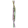
Mass: 200.590 Da / Num. of mol.: 2 / Source method: obtained synthetically / Formula: Hg
#3: Water ChemComp-HOH / water


Mass: 18.015 Da / Num. of mol.: 123 / Source method: isolated from a natural source / Formula: H2O

-
Experimental details

-
Experiment

ExperimentMethod: X-RAY DIFFRACTION / Number of used crystals: 1

-
Sample preparation

CrystalDensity Matthews: 2.11 Å3/Da / Density % sol: 41.32 % / Description: NONE
Crystal growpH: 6.8
Details: 4% PEG 8000, 50 MM SODIUM PHOSPHATE PH 6.8, 100 MM NACL. PROTEIN CONCENTRATION 20 MG/ML. CRYOBUFFER = RESERVOIR PLUS 17% PEG 400.

-
Data collection

DiffractionMean temperature: 100 K
Diffraction sourceSource: SYNCHROTRON / Site: ESRF / Beamline: ID14-1 / Wavelength: 0.933
DetectorType: ADSC CCD / Detector: CCD / Date: Feb 24, 2005
RadiationProtocol: SINGLE WAVELENGTH / Monochromatic (M) / Laue (L): M / Scattering type: x-ray
Radiation wavelengthWavelength: 0.933 Å / Relative weight: 1
ReflectionResolution: 2.1→49.3 Å / Num. obs: 300363 / % possible obs: 99.8 % / Observed criterion σ(I): 2 / Redundancy: 11.5 % / Rmerge(I) obs: 0.08 / Net I/σ(I): 5
Reflection shellResolution: 2.1→2.17 Å / Redundancy: 12 % / Rmerge(I) obs: 0.31 / Mean I/σ(I) obs: 2.2 / % possible all: 100

-
Processing

Software
NameVersionClassification
REFMAC5.2.0005refinement
MOSFLMdata reduction
SCALAdata scaling
SHARPphasing
RefinementMethod to determine structure: MAD
Starting model: NONE

Resolution: 2.1→18 Å / Cor.coef. Fo:Fc: 0.931 / Cor.coef. Fo:Fc free: 0.876 / SU B: 14.284 / SU ML: 0.192 / TLS residual ADP flag: LIKELY RESIDUAL / Cross valid method: THROUGHOUT / ESU R: 0.312 / ESU R Free: 0.258 / Stereochemistry target values: MAXIMUM LIKELIHOOD / Details: HYDROGENS HAVE BEEN ADDED IN THE RIDING POSITIONS.
RfactorNum. reflection% reflectionSelection details
Rfree0.31 1074 4.1 %RANDOM
Rwork0.233 ---
obs0.236 24984 99.9 %-
Solvent computationIon probe radii: 0.8 Å / Shrinkage radii: 0.8 Å / VDW probe radii: 1.2 Å / Solvent model: BABINET MODEL WITH MASK
Displacement parametersBiso mean: 30.26 Å2
Baniso -1Baniso -2Baniso -3
1--3.52 Å20 Å2-2.25 Å2
2--2.86 Å20 Å2
3----0.54 Å2
Refinement stepCycle: LAST / Resolution: 2.1→18 Å
ProteinNucleic acidLigandSolventTotal
Num. atoms3700 0 2 123 3825
Refine LS restraints
Refine-IDTypeDev idealDev ideal targetNumber
X-RAY DIFFRACTIONr_bond_refined_d0.0260.0223728
X-RAY DIFFRACTIONr_bond_other_d
X-RAY DIFFRACTIONr_angle_refined_deg1.9221.9685000
X-RAY DIFFRACTIONr_angle_other_deg
X-RAY DIFFRACTIONr_dihedral_angle_1_deg5.7315440
X-RAY DIFFRACTIONr_dihedral_angle_2_deg39.38625.593236
X-RAY DIFFRACTIONr_dihedral_angle_3_deg19.63115760
X-RAY DIFFRACTIONr_dihedral_angle_4_deg19.6271536
X-RAY DIFFRACTIONr_chiral_restr0.1350.2552
X-RAY DIFFRACTIONr_gen_planes_refined0.0080.022844
X-RAY DIFFRACTIONr_gen_planes_other
X-RAY DIFFRACTIONr_nbd_refined0.2480.21777
X-RAY DIFFRACTIONr_nbd_other
X-RAY DIFFRACTIONr_nbtor_refined0.2990.22577
X-RAY DIFFRACTIONr_nbtor_other
X-RAY DIFFRACTIONr_xyhbond_nbd_refined0.240.2147
X-RAY DIFFRACTIONr_xyhbond_nbd_other
X-RAY DIFFRACTIONr_metal_ion_refined
X-RAY DIFFRACTIONr_metal_ion_other
X-RAY DIFFRACTIONr_symmetry_vdw_refined0.340.2101
X-RAY DIFFRACTIONr_symmetry_vdw_other
X-RAY DIFFRACTIONr_symmetry_hbond_refined0.2760.224
X-RAY DIFFRACTIONr_symmetry_hbond_other
X-RAY DIFFRACTIONr_symmetry_metal_ion_refined
X-RAY DIFFRACTIONr_symmetry_metal_ion_other
X-RAY DIFFRACTIONr_mcbond_it1.2791.52341
X-RAY DIFFRACTIONr_mcbond_other
X-RAY DIFFRACTIONr_mcangle_it1.83623548
X-RAY DIFFRACTIONr_mcangle_other
X-RAY DIFFRACTIONr_scbond_it3.48631590
X-RAY DIFFRACTIONr_scbond_other
X-RAY DIFFRACTIONr_scangle_it5.3354.51452
X-RAY DIFFRACTIONr_scangle_other
X-RAY DIFFRACTIONr_long_range_B_refined
X-RAY DIFFRACTIONr_long_range_B_other
X-RAY DIFFRACTIONr_rigid_bond_restr
X-RAY DIFFRACTIONr_sphericity_free
X-RAY DIFFRACTIONr_sphericity_bonded
LS refinement shellResolution: 2.1→2.15 Å / Total num. of bins used: 20 /
RfactorNum. reflection
Rfree0.344 90
Rwork0.233 1794
Refinement TLS params.

Method: refined / Refine-ID: X-RAY DIFFRACTION

IDL112)L122)L132)L222)L232)L332)S11 (Å °)S12 (Å °)S13 (Å °)S21 (Å °)S22 (Å °)S23 (Å °)S31 (Å °)S32 (Å °)S33 (Å °)T112)T122)T132)T222)T232)T332)Origin x (Å)Origin y (Å)Origin z (Å)
111.7525-1.51376.94190.2504-0.92644.11920.1331-0.219-0.1078-0.0215-0.04110.00830.1288-0.1414-0.092-0.04080.03330.0458-0.0102-0.00610.0073-18.6731.76562.433
214.0578-2.82829.3490.569-1.88096.2175-0.0940.17660.21880.0062-0.0436-0.0581-0.04850.1520.13760.00570.03630.0193-0.00660.0260.0081-14.73132.64459.192
317.4883-2.52689.54690.4949-1.45565.2767-0.35380.26790.26660.04650.13130.0407-0.25790.09740.22260.0018-0.00650.01820.0017-0.00580.0073-64.34842.87434.333
49.0595-1.61896.06130.4356-1.20624.159-0.0526-0.75080.1913-0.03230.11660.05670.0429-0.5046-0.06390.00670.03370.0437-0.010.00940.0161-64.2239.24831.017
Refinement TLS group
IDRefine-IDRefine TLS-IDAuth asym-IDAuth seq-ID
1X-RAY DIFFRACTION1E58 - 168
2X-RAY DIFFRACTION2B58 - 168
3X-RAY DIFFRACTION3C58 - 168
4X-RAY DIFFRACTION4D58 - 168

+
About Yorodumi

-
News

-
Feb 9, 2022. New format data for meta-information of EMDB entries

New format data for meta-information of EMDB entries

  • Version 3 of the EMDB header file is now the official format.
  • The previous official version 1.9 will be removed from the archive.

Related info.:EMDB header

External links:wwPDB to switch to version 3 of the EMDB data model

-
Aug 12, 2020. Covid-19 info

Covid-19 info

URL: https://pdbjlc1.pdbj.org/emnavi/covid19.php

New page: Covid-19 featured information page in EM Navigator.

Related info.:Covid-19 info / Mar 5, 2020. Novel coronavirus structure data

+
Mar 5, 2020. Novel coronavirus structure data

Novel coronavirus structure data

Related info.:Yorodumi Speices / Aug 12, 2020. Covid-19 info

External links:COVID-19 featured content - PDBj / Molecule of the Month (242):Coronavirus Proteases

+
Jan 31, 2019. EMDB accession codes are about to change! (news from PDBe EMDB page)

EMDB accession codes are about to change! (news from PDBe EMDB page)

  • The allocation of 4 digits for EMDB accession codes will soon come to an end. Whilst these codes will remain in use, new EMDB accession codes will include an additional digit and will expand incrementally as the available range of codes is exhausted. The current 4-digit format prefixed with “EMD-” (i.e. EMD-XXXX) will advance to a 5-digit format (i.e. EMD-XXXXX), and so on. It is currently estimated that the 4-digit codes will be depleted around Spring 2019, at which point the 5-digit format will come into force.
  • The EM Navigator/Yorodumi systems omit the EMD- prefix.

Related info.:Q: What is EMD? / ID/Accession-code notation in Yorodumi/EM Navigator

External links:EMDB Accession Codes are Changing Soon! / Contact to PDBj

+
Jul 12, 2017. Major update of PDB

Major update of PDB

  • wwPDB released updated PDB data conforming to the new PDBx/mmCIF dictionary.
  • This is a major update changing the version number from 4 to 5, and with Remediation, in which all the entries are updated.
  • In this update, many items about electron microscopy experimental information are reorganized (e.g. em_software).
  • Now, EM Navigator and Yorodumi are based on the updated data.

External links:wwPDB Remediation / Enriched Model Files Conforming to OneDep Data Standards Now Available in the PDB FTP Archive

-
Yorodumi

Thousand views of thousand structures

  • Yorodumi is a browser for structure data from EMDB, PDB, SASBDB, etc.
  • This page is also the successor to EM Navigator detail page, and also detail information page/front-end page for Omokage search.
  • The word "yorodu" (or yorozu) is an old Japanese word meaning "ten thousand". "mi" (miru) is to see.

Related info.:EMDB / PDB / SASBDB / Comparison of 3 databanks / Yorodumi Search / Aug 31, 2016. New EM Navigator & Yorodumi / Yorodumi Papers / Jmol/JSmol / Function and homology information / Changes in new EM Navigator and Yorodumi

Read more