 Movie
Movie Controller

[English] 日本語
 Yorodumi
- PDB-2v4c: Structure of sialic acid binding protein (SiaP) in the presence of KDN -
+ Open data

- Basic information
| Entry | Database: PDB / ID: 2v4c | |||||||||
|---|---|---|---|---|---|---|---|---|---|---|
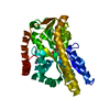
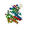
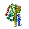
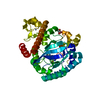
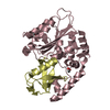
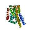
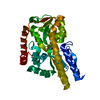
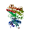
| Title | Structure of sialic acid binding protein (SiaP) in the presence of KDN | |||||||||
 Components Components | SIALIC ACID-BINDING PERIPLASMIC PROTEIN SIAP | |||||||||
 Keywords Keywords | TRANSPORT PROTEIN / SUGAR TRANSPORT / TRANSPORT PROTEIN ESR / TRAP | |||||||||
| Function / homology |  Function and homology information Function and homology information | |||||||||
| Biological species |  HAEMOPHILUS INFLUENZAE (bacteria) HAEMOPHILUS INFLUENZAE (bacteria) | |||||||||
| Method |  X-RAY DIFFRACTION / SYNCHROTRON / MOLECULAR REPLACEMENT / Resolution: 1.7 Å X-RAY DIFFRACTION / SYNCHROTRON / MOLECULAR REPLACEMENT / Resolution: 1.7 Å | |||||||||
 Authors Authors | Fischer, M. / Hubbard, R.E. | |||||||||
 Citation Citation | Journal: J.Am.Chem.Soc. / Year: 2019 Title: Water networks can determine the affinity of ligand binding to proteins. Authors: Darby, J.F. / Hopkins, A.P. / Shimizu, S. / Roberts, S.M. / Brannigan, J.A. / Turkenburg, J.P. / Thomas, G.H. / Hubbard, R.E. / Fischer, M. | |||||||||
| History |
|
- Structure visualization
| Structure viewer | Molecule: MolmilJmol/JSmol |
|---|
- Downloads & links
-Download
| PDBx/mmCIF format | 2v4c.cif.gz | 148.2 KB | Display | PDBx/mmCIF format |
|---|---|---|---|---|
| PDB format | pdb2v4c.ent.gz | 115.8 KB | Display | PDB format |
| PDBx/mmJSON format | 2v4c.json.gz | Tree view | PDBx/mmJSON format | |
| Others |  Other downloads Other downloads |
-Validation report
| Arichive directory | https://data.pdbj.org/pub/pdb/validation_reports/v4/2v4cftp://data.pdbj.org/pub/pdb/validation_reports/v4/2v4c | HTTPS FTP |
|---|
-Related structure data
| Related structure data |  2wx9C  2wykC  2wypC  2xa5C 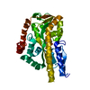 6h75C 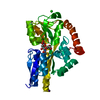 6h76C  3b50S C: citing same article ( S: Starting model for refinement |
|---|---|
| Similar structure data |


-Links
 PDBj
PDBj- Assembly
| Deposited unit | 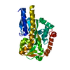
| ||||||||
|---|---|---|---|---|---|---|---|---|---|
| 1 |
| ||||||||
| Unit cell |
|
-Components
| #1: Protein | Mass: 34618.184 Da / Num. of mol.: 1 Source method: isolated from a genetically manipulated source Source: (gene. exp.) HAEMOPHILUS INFLUENZAE (bacteria) / Plasmid: PET21B / Production host: |
|---|---|
| #2: Sugar | ChemComp-KDN /   Type: D-saccharide, beta linking / Mass: 268.218 Da / Num. of mol.: 1 Type: D-saccharide, beta linking / Mass: 268.218 Da / Num. of mol.: 1Source method: isolated from a genetically manipulated source Formula: C9H16O9 |
| #3: Water | ChemComp-HOH /  Mass: 18.015 Da / Num. of mol.: 346 / Source method: isolated from a natural source / Formula: H2O Mass: 18.015 Da / Num. of mol.: 346 / Source method: isolated from a natural source / Formula: H2O |
-Experimental details
-Experiment
| Experiment | Method: X-RAY DIFFRACTION / Number of used crystals: 1 |
|---|
- Sample preparation
| Crystal | Density Matthews: 2.18 Å3/Da / Density % sol: 43.6 % / Description: NONE |
|---|---|
| Crystal grow | Temperature: 277 K / pH: 7 Details: 36 MG/ML SIAP (IN 20 MM HEPES, 10 MM NACL, 10 MM KDN, PH 8.0) AND EQUAL VOLUME OF RESERVOIR SOLUTION (100MM MES PH 6.0 28.5% PEG 6K); TEMPERATURE 277K |
-Data collection
| Diffraction | Mean temperature: 100 K |
|---|---|
| Diffraction source | Source: SYNCHROTRON / Site: ESRF  / Beamline: ID29 / Wavelength: 0.9755 / Beamline: ID29 / Wavelength: 0.9755 |
| Detector | Type: ADSC CCD / Detector: CCD / Date: Jul 2, 2008 |
| Radiation | Protocol: SINGLE WAVELENGTH / Monochromatic (M) / Laue (L): M / Scattering type: x-ray |
| Radiation wavelength | Wavelength: 0.9755 Å / Relative weight: 1 |
| Reflection | Resolution: 1.7→50 Å / Num. obs: 34122 / % possible obs: 99.2 % / Redundancy: 6.5 % / Rmerge(I) obs: 0.09 / Net I/σ(I): 17.5 |
| Reflection shell | Resolution: 1.7→1.73 Å / Redundancy: 5.2 % / Rmerge(I) obs: 0.44 / Mean I/σ(I) obs: 2.43 / % possible all: 93.9 |
- Processing
| Software |
| ||||||||||||||||||||||||||||||||||||||||||||||||||||||||||||||||||||||||||||||||||||||||||||||||||||||||||||||||||||||||||||||||||||||||||||||||||||||||||||||||||||||||||||||||||||||
|---|---|---|---|---|---|---|---|---|---|---|---|---|---|---|---|---|---|---|---|---|---|---|---|---|---|---|---|---|---|---|---|---|---|---|---|---|---|---|---|---|---|---|---|---|---|---|---|---|---|---|---|---|---|---|---|---|---|---|---|---|---|---|---|---|---|---|---|---|---|---|---|---|---|---|---|---|---|---|---|---|---|---|---|---|---|---|---|---|---|---|---|---|---|---|---|---|---|---|---|---|---|---|---|---|---|---|---|---|---|---|---|---|---|---|---|---|---|---|---|---|---|---|---|---|---|---|---|---|---|---|---|---|---|---|---|---|---|---|---|---|---|---|---|---|---|---|---|---|---|---|---|---|---|---|---|---|---|---|---|---|---|---|---|---|---|---|---|---|---|---|---|---|---|---|---|---|---|---|---|---|---|---|---|
| Refinement | Method to determine structure: MOLECULAR REPLACEMENT Starting model: PDB ENTRY 3B50 Resolution: 1.7→41.56 Å / Cor.coef. Fo:Fc: 0.973 / Cor.coef. Fo:Fc free: 0.957 / SU B: 4.03 / SU ML: 0.06 / Cross valid method: THROUGHOUT / ESU R: 0.163 / ESU R Free: 0.1 / Stereochemistry target values: MAXIMUM LIKELIHOOD Details: HYDROGENS HAVE BEEN ADDED IN THE RIDING POSITIONS.U VALUES REFINED INDIVIDUALLY.
| ||||||||||||||||||||||||||||||||||||||||||||||||||||||||||||||||||||||||||||||||||||||||||||||||||||||||||||||||||||||||||||||||||||||||||||||||||||||||||||||||||||||||||||||||||||||
| Solvent computation | Ion probe radii: 0.8 Å / Shrinkage radii: 0.8 Å / VDW probe radii: 1.2 Å / Solvent model: MASK | ||||||||||||||||||||||||||||||||||||||||||||||||||||||||||||||||||||||||||||||||||||||||||||||||||||||||||||||||||||||||||||||||||||||||||||||||||||||||||||||||||||||||||||||||||||||
| Displacement parameters | Biso mean: 21.302 Å2
| ||||||||||||||||||||||||||||||||||||||||||||||||||||||||||||||||||||||||||||||||||||||||||||||||||||||||||||||||||||||||||||||||||||||||||||||||||||||||||||||||||||||||||||||||||||||
| Refinement step | Cycle: LAST / Resolution: 1.7→41.56 Å
| ||||||||||||||||||||||||||||||||||||||||||||||||||||||||||||||||||||||||||||||||||||||||||||||||||||||||||||||||||||||||||||||||||||||||||||||||||||||||||||||||||||||||||||||||||||||
| Refine LS restraints |
|