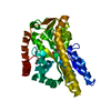
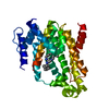
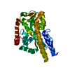
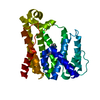
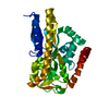
[English] 日本語
Yorodumi
- PDB-2xwo: SiaP R147E mutant in complex with sialylamide -

+
Open data


ID or keywords:

Loading...

-
Basic information

Entry
Database: PDB / ID: 2xwo
TitleSiaP R147E mutant in complex with sialylamide
ComponentsSIALIC ACID-BINDING PERIPLASMIC PROTEIN SIAP
KeywordsTRANSPORT PROTEIN / TRAP / SUGAR TRANSPORT
Function / homology
Function and homology information


: / transmembrane transport / outer membrane-bounded periplasmic space
Similarity search - Function
TRAP transporter solute receptor, DctP family / Bacterial extracellular solute-binding protein, family 7 / TRAP transporter solute receptor DctP / TRAP transporter solute receptor DctP superfamily / Bacterial extracellular solute-binding protein, family 7 / D-Maltodextrin-Binding Protein; domain 2 / 3-Layer(aba) Sandwich / Alpha Beta
Similarity search - Domain/homology
Chem-SLM / Sialic acid-binding periplasmic protein SiaP
Similarity search - Component
Biological speciesHAEMOPHILUS INFLUENZAE (bacteria)
MethodX-RAY DIFFRACTION / SYNCHROTRON / MOLECULAR REPLACEMENT / Resolution: 1.54 Å
AuthorsFischer, M. / Hubbard, R.E.
CitationJournal: J.Biol.Chem. / Year: 2015
Title: Tripartite ATP-Independent Periplasmic (Trap) Transporters Use an Arginine-Mediated Selectivity Filter for High Affinity Substrate Binding.
Authors: Fischer, M. / Hopkins, A.P. / Severi, E. / Hawkhead, J. / Bawdon, D. / Watts, A.G. / Hubbard, R.E. / Thomas, G.H.
History
DepositionNov 4, 2010Deposition site: PDBE / Processing site: PDBE
Revision 1.0Nov 16, 2011Provider: repository / Type: Initial release
Revision 1.1Sep 16, 2015Group: Database references
Revision 1.2Sep 23, 2015Group: Database references
Revision 1.3Nov 18, 2015Group: Database references
Revision 1.4Mar 6, 2019Group: Data collection / Experimental preparation / Other
Category: exptl_crystal_grow / pdbx_database_proc / pdbx_database_status
Item: _exptl_crystal_grow.temp / _pdbx_database_status.recvd_author_approval
Revision 1.5Jul 29, 2020Group: Derived calculations / Other / Structure summary
Category: chem_comp / entity ...chem_comp / entity / pdbx_database_status / pdbx_entity_nonpoly / struct_site / struct_site_gen
Item: _chem_comp.name / _chem_comp.type ..._chem_comp.name / _chem_comp.type / _entity.pdbx_description / _pdbx_database_status.status_code_sf / _pdbx_entity_nonpoly.name
Description: Carbohydrate remediation / Provider: repository / Type: Remediation
Revision 1.6Dec 20, 2023Group: Data collection / Database references ...Data collection / Database references / Refinement description / Structure summary
Category: chem_comp / chem_comp_atom ...chem_comp / chem_comp_atom / chem_comp_bond / database_2 / pdbx_initial_refinement_model
Item: _chem_comp.pdbx_synonyms / _database_2.pdbx_DOI / _database_2.pdbx_database_accession

-
Structure visualization

Structure viewerMolecule:
MolmilJmol/JSmol

Downloads & links

-
Assembly

Deposited unit
A: SIALIC ACID-BINDING PERIPLASMIC PROTEIN SIAP
hetero molecules


Theoretical massNumber of molelcules
Total (without water)35,3132
Polymers35,0051
Non-polymers3081
Water7,008389
1


  • Idetical with deposited unit
  • defined by author&software
TypeNameSymmetry operationNumber
identity operation1_555x,y,z1
MethodPISA
Unit cell
Length a, b, c (Å)47.860, 74.760, 87.170
Angle α, β, γ (deg.)90.00, 90.00, 90.00
Int Tables number19
Space group name H-MP212121

-
Components

#1: Protein SIALIC ACID-BINDING PERIPLASMIC PROTEIN SIAP / EXTRACYTOPLASMIC SOLUTE RECEPTOR PROTEIN SIAP / N-ACETYLNEURAMINIC-BINDING PROTEIN / NEU5AC-BINDING ...EXTRACYTOPLASMIC SOLUTE RECEPTOR PROTEIN SIAP / N-ACETYLNEURAMINIC-BINDING PROTEIN / NEU5AC-BINDING PROTEIN / SUBSTRATE BINDING PROTEIN


Mass: 35004.539 Da / Num. of mol.: 1 / Mutation: YES
Source method: isolated from a genetically manipulated source
Source: (gene. exp.) HAEMOPHILUS INFLUENZAE (bacteria) / Production host: ESCHERICHIA COLI (E. coli) / Strain (production host): BL21(DE3) / Variant (production host): PLYSS PAH16 / References: UniProt: P44542
#2: Sugar ChemComp-SLM / (2S,4S,5R,6R)-5-acetamido-2,4-dihydroxy-6-[(1R,2R)-1,2,3-trihydroxypropyl]oxane-2-carboxamide / SIALYLAMIDE


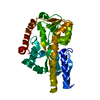
Type: D-saccharide, beta linking / Mass: 308.285 Da / Num. of mol.: 1 / Source method: obtained synthetically / Formula: C11H20N2O8
#3: Water ChemComp-HOH / water


Mass: 18.015 Da / Num. of mol.: 389 / Source method: isolated from a natural source / Formula: H2O
Compound detailsENGINEERED RESIDUE IN CHAIN A, ARG 170 TO GLU

-
Experimental details

-
Experiment

ExperimentMethod: X-RAY DIFFRACTION / Number of used crystals: 1

-
Sample preparation

CrystalDensity Matthews: 2.2 Å3/Da / Density % sol: 45 % / Description: NONE
Crystal growTemperature: 277 K / pH: 7
Details: 35 MG/ML SIAP (IN 20 MM HEPES, 10 MM NACL, 20 MM SIALYLAMIDE, PH 8.0) WITH AN EQUIVOLUME OF RESERVOIR SOLUTION (100MM MES PH 6.0 28.5% PEG 6K, 2% (W/V) BENZAMIDINE HYDROCHLORIDE HYDRATE); TEMPERATURE 277K

-
Data collection

DiffractionMean temperature: 100 K
Diffraction sourceSource: SYNCHROTRON / Site: ESRF / Beamline: ID23-2 / Wavelength: 0.8726
DetectorType: ADSC CCD / Detector: CCD / Date: Oct 26, 2009
RadiationProtocol: SINGLE WAVELENGTH / Monochromatic (M) / Laue (L): M / Scattering type: x-ray
Radiation wavelengthWavelength: 0.8726 Å / Relative weight: 1
ReflectionResolution: 1.54→29.06 Å / Num. obs: 47100 / % possible obs: 100 % / Observed criterion σ(I): 2.5 / Redundancy: 7.3 % / Biso Wilson estimate: 14.4 Å2 / Rmerge(I) obs: 0.12 / Net I/σ(I): 11.9
Reflection shellResolution: 1.54→1.62 Å / Redundancy: 7.3 % / Rmerge(I) obs: 0.97 / Mean I/σ(I) obs: 2.5 / % possible all: 100

-
Processing

Software
NameVersionClassification
REFMAC5.5.0109refinement
MOSFLMdata reduction
SCALEPACKdata scaling
REFMACRIGID BODYphasing
RefinementMethod to determine structure: MOLECULAR REPLACEMENT
Starting model: PDB ENTRY 2V4C
Resolution: 1.54→28.37 Å / Cor.coef. Fo:Fc: 0.976 / Cor.coef. Fo:Fc free: 0.962 / SU B: 2.911 / SU ML: 0.047 / Cross valid method: THROUGHOUT / ESU R: 0.081 / ESU R Free: 0.072 / Stereochemistry target values: MAXIMUM LIKELIHOOD
Details: HYDROGENS HAVE BEEN ADDED IN THE RIDING POSITIONS. U VALUES REFINED INDIVIDUALLY
RfactorNum. reflection% reflectionSelection details
Rfree0.17891 2384 5.1 %RANDOM
Rwork0.13037 ---
obs0.13287 44650 100 %-
Solvent computationIon probe radii: 0.8 Å / Shrinkage radii: 0.8 Å / VDW probe radii: 1.4 Å / Solvent model: BABINET MODEL WITH MASK
Displacement parametersBiso mean: 14.015 Å2
Baniso -1Baniso -2Baniso -3
1--0.28 Å20 Å20 Å2
2---0.48 Å20 Å2
3---0.77 Å2
Refinement stepCycle: LAST / Resolution: 1.54→28.37 Å
ProteinNucleic acidLigandSolventTotal
Num. atoms2426 0 21 389 2836
Refine LS restraints
Refine-IDTypeDev idealDev ideal targetNumber
X-RAY DIFFRACTIONr_bond_refined_d0.0170.0222584
X-RAY DIFFRACTIONr_bond_other_d0.0020.021728
X-RAY DIFFRACTIONr_angle_refined_deg1.5551.9743511
X-RAY DIFFRACTIONr_angle_other_deg1.0073.0024256
X-RAY DIFFRACTIONr_dihedral_angle_1_deg5.7035330
X-RAY DIFFRACTIONr_dihedral_angle_2_deg39.19626.23122
X-RAY DIFFRACTIONr_dihedral_angle_3_deg13.64815460
X-RAY DIFFRACTIONr_dihedral_angle_4_deg14.963154
X-RAY DIFFRACTIONr_chiral_restr0.1010.2387
X-RAY DIFFRACTIONr_gen_planes_refined0.0080.0212914
X-RAY DIFFRACTIONr_gen_planes_other0.0010.02490
X-RAY DIFFRACTIONr_nbd_refined
X-RAY DIFFRACTIONr_nbd_other
X-RAY DIFFRACTIONr_nbtor_refined
X-RAY DIFFRACTIONr_nbtor_other
X-RAY DIFFRACTIONr_xyhbond_nbd_refined
X-RAY DIFFRACTIONr_xyhbond_nbd_other
X-RAY DIFFRACTIONr_metal_ion_refined
X-RAY DIFFRACTIONr_metal_ion_other
X-RAY DIFFRACTIONr_symmetry_vdw_refined
X-RAY DIFFRACTIONr_symmetry_vdw_other
X-RAY DIFFRACTIONr_symmetry_hbond_refined
X-RAY DIFFRACTIONr_symmetry_hbond_other
X-RAY DIFFRACTIONr_symmetry_metal_ion_refined
X-RAY DIFFRACTIONr_symmetry_metal_ion_other
X-RAY DIFFRACTIONr_mcbond_it1.5251.51592
X-RAY DIFFRACTIONr_mcbond_other0.5581.5637
X-RAY DIFFRACTIONr_mcangle_it2.31622569
X-RAY DIFFRACTIONr_mcangle_other
X-RAY DIFFRACTIONr_scbond_it3.7783992
X-RAY DIFFRACTIONr_scbond_other
X-RAY DIFFRACTIONr_scangle_it5.4694.5937
X-RAY DIFFRACTIONr_scangle_other
X-RAY DIFFRACTIONr_long_range_B_refined
X-RAY DIFFRACTIONr_long_range_B_other
X-RAY DIFFRACTIONr_rigid_bond_restr1.56534312
X-RAY DIFFRACTIONr_sphericity_free
X-RAY DIFFRACTIONr_sphericity_bonded
LS refinement shellResolution: 1.54→1.58 Å / Total num. of bins used: 20
RfactorNum. reflection% reflection
Rfree0.275 178 -
Rwork0.207 3235 -
obs--99.97 %

+
About Yorodumi

-
News

-
Feb 9, 2022. New format data for meta-information of EMDB entries

New format data for meta-information of EMDB entries

  • Version 3 of the EMDB header file is now the official format.
  • The previous official version 1.9 will be removed from the archive.

Related info.:EMDB header

External links:wwPDB to switch to version 3 of the EMDB data model

-
Aug 12, 2020. Covid-19 info

Covid-19 info

URL: https://pdbjlvh1.pdbj.org/emnavi/covid19.php

New page: Covid-19 featured information page in EM Navigator.

Related info.:Covid-19 info / Mar 5, 2020. Novel coronavirus structure data

+
Mar 5, 2020. Novel coronavirus structure data

Novel coronavirus structure data

Related info.:Yorodumi Speices / Aug 12, 2020. Covid-19 info

External links:COVID-19 featured content - PDBj / Molecule of the Month (242):Coronavirus Proteases

+
Jan 31, 2019. EMDB accession codes are about to change! (news from PDBe EMDB page)

EMDB accession codes are about to change! (news from PDBe EMDB page)

  • The allocation of 4 digits for EMDB accession codes will soon come to an end. Whilst these codes will remain in use, new EMDB accession codes will include an additional digit and will expand incrementally as the available range of codes is exhausted. The current 4-digit format prefixed with “EMD-” (i.e. EMD-XXXX) will advance to a 5-digit format (i.e. EMD-XXXXX), and so on. It is currently estimated that the 4-digit codes will be depleted around Spring 2019, at which point the 5-digit format will come into force.
  • The EM Navigator/Yorodumi systems omit the EMD- prefix.

Related info.:Q: What is EMD? / ID/Accession-code notation in Yorodumi/EM Navigator

External links:EMDB Accession Codes are Changing Soon! / Contact to PDBj

+
Jul 12, 2017. Major update of PDB

Major update of PDB

  • wwPDB released updated PDB data conforming to the new PDBx/mmCIF dictionary.
  • This is a major update changing the version number from 4 to 5, and with Remediation, in which all the entries are updated.
  • In this update, many items about electron microscopy experimental information are reorganized (e.g. em_software).
  • Now, EM Navigator and Yorodumi are based on the updated data.

External links:wwPDB Remediation / Enriched Model Files Conforming to OneDep Data Standards Now Available in the PDB FTP Archive

-
Yorodumi

Thousand views of thousand structures

  • Yorodumi is a browser for structure data from EMDB, PDB, SASBDB, etc.
  • This page is also the successor to EM Navigator detail page, and also detail information page/front-end page for Omokage search.
  • The word "yorodu" (or yorozu) is an old Japanese word meaning "ten thousand". "mi" (miru) is to see.

Related info.:EMDB / PDB / SASBDB / Comparison of 3 databanks / Yorodumi Search / Aug 31, 2016. New EM Navigator & Yorodumi / Yorodumi Papers / Jmol/JSmol / Function and homology information / Changes in new EM Navigator and Yorodumi

Read more