 Movie
Movie Controller

[English] 日本語
 Yorodumi
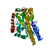
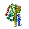
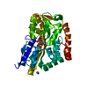
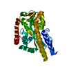
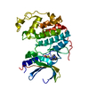
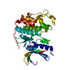
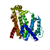
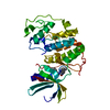
- PDB-4mmp: Structure of Sialic Acid Binding Protein from Pasturella Multocida -
+ Open data

- Basic information
| Entry | Database: PDB / ID: 4mmp | ||||||
|---|---|---|---|---|---|---|---|
| Title | Structure of Sialic Acid Binding Protein from Pasturella Multocida | ||||||
 Components Components | Sialic Acid Binding Protein | ||||||
 Keywords Keywords | SUGAR BINDING PROTEIN / Sugar Transport / TRAP Transporter | ||||||
| Function / homology | Bacterial extracellular solute-binding protein, family 7 / D-Maltodextrin-Binding Protein; domain 2 / 3-Layer(aba) Sandwich / Alpha Beta / N-acetyl-beta-neuraminic acid / :  Function and homology information Function and homology information | ||||||
| Biological species |  Pasteurella multocida subsp. gallicida P1059 (bacteria) Pasteurella multocida subsp. gallicida P1059 (bacteria) | ||||||
| Method |  X-RAY DIFFRACTION / SYNCHROTRON / MOLECULAR REPLACEMENT / molecular replacement / Resolution: 1.57 Å X-RAY DIFFRACTION / SYNCHROTRON / MOLECULAR REPLACEMENT / molecular replacement / Resolution: 1.57 Å | ||||||
 Authors Authors | Ramaswamy, S. / Thanuja, G.S. | ||||||
 Citation Citation | Journal: Acta Crystallogr.,Sect.D / Year: 2014 Title: Bacterial periplasmic sialic acid-binding proteins exhibit a conserved binding site. Authors: Gangi Setty, T. / Cho, C. / Govindappa, S. / Apicella, M.A. / Ramaswamy, S. #1: Journal: J.Biol.Chem. / Year: 2008Title: Characterization of the N-acetyl-5-neuraminic acid-binding site of the extracytoplasmic solute receptor (SiaP) of nontypeable Haemophilus influenzae strain 2019. Authors: Johnston, J.W. / Coussens, N.P. / Allen, S. / Houtman, J.C. / Turner, K.H. / Zaleski, A. / Ramaswamy, S. / Gibson, B.W. / Apicella, M.A. | ||||||
| History |
|
- Structure visualization
| Structure viewer | Molecule: MolmilJmol/JSmol |
|---|
- Downloads & links
-Download
| PDBx/mmCIF format | 4mmp.cif.gz | 78.2 KB | Display | PDBx/mmCIF format |
|---|---|---|---|---|
| PDB format | pdb4mmp.ent.gz | 57.3 KB | Display | PDB format |
| PDBx/mmJSON format | 4mmp.json.gz | Tree view | PDBx/mmJSON format | |
| Others |  Other downloads Other downloads |
-Validation report
| Arichive directory | https://data.pdbj.org/pub/pdb/validation_reports/mm/4mmpftp://data.pdbj.org/pub/pdb/validation_reports/mm/4mmp | HTTPS FTP |
|---|
-Related structure data
| Related structure data | 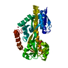 4magC 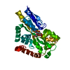 4mnpC  3b50S S: Starting model for refinement C: citing same article ( |
|---|---|
| Similar structure data |


-Links
 PDBj
PDBj- Assembly
| Deposited unit | 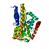
| ||||||||
|---|---|---|---|---|---|---|---|---|---|
| 1 |
| ||||||||
| Unit cell |
|
-Components
| #1: Protein | Mass: 34206.879 Da / Num. of mol.: 1 / Fragment: UNP residues 22-327 Source method: isolated from a genetically manipulated source Source: (gene. exp.) Pasteurella multocida subsp. gallicida P1059 (bacteria)Gene: SiaP, P1059_01877 / Plasmid: pET28A / Production host: |
|---|---|
| #2: Sugar | ChemComp-SLB / 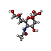  Type: D-saccharide, beta linking / Mass: 309.270 Da / Num. of mol.: 1 Type: D-saccharide, beta linking / Mass: 309.270 Da / Num. of mol.: 1Source method: isolated from a genetically manipulated source Formula: C11H19NO9 |
| #3: Water | ChemComp-HOH /  Mass: 18.015 Da / Num. of mol.: 193 / Source method: isolated from a natural source / Formula: H2O Mass: 18.015 Da / Num. of mol.: 193 / Source method: isolated from a natural source / Formula: H2O |
-Experimental details
-Experiment
| Experiment | Method: X-RAY DIFFRACTION / Number of used crystals: 1 |
|---|
- Sample preparation
| Crystal | Density Matthews: 2.07 Å3/Da / Density % sol: 40.65 % |
|---|---|
| Crystal grow | Temperature: 277 K / Method: vapor diffusion, hanging drop / pH: 6.5 Details: Drops were setup with equal volume of protein and 1.6M Sodium citrate tribasic dihydrate pH 6.5(crystallization buffer) and suspended over 100 l of crystallization buffer, VAPOR DIFFUSION, ...Details: Drops were setup with equal volume of protein and 1.6M Sodium citrate tribasic dihydrate pH 6.5(crystallization buffer) and suspended over 100 l of crystallization buffer, VAPOR DIFFUSION, HANGING DROP, temperature 277K |
-Data collection
| Diffraction | Mean temperature: 100 K | ||||||||||||||||||||||||||||||||||||||||||||||||||||||||||||||||||||||||||||||||||||||||
|---|---|---|---|---|---|---|---|---|---|---|---|---|---|---|---|---|---|---|---|---|---|---|---|---|---|---|---|---|---|---|---|---|---|---|---|---|---|---|---|---|---|---|---|---|---|---|---|---|---|---|---|---|---|---|---|---|---|---|---|---|---|---|---|---|---|---|---|---|---|---|---|---|---|---|---|---|---|---|---|---|---|---|---|---|---|---|---|---|---|
| Diffraction source | Source: SYNCHROTRON / Site: APS  / Beamline: 17-ID / Wavelength: 1 Å / Beamline: 17-ID / Wavelength: 1 Å | ||||||||||||||||||||||||||||||||||||||||||||||||||||||||||||||||||||||||||||||||||||||||
| Detector | Type: DECTRIS PILATUS 6M / Detector: PIXEL / Date: Feb 21, 2012 / Details: mirrors | ||||||||||||||||||||||||||||||||||||||||||||||||||||||||||||||||||||||||||||||||||||||||
| Radiation | Protocol: SINGLE WAVELENGTH / Monochromatic (M) / Laue (L): M / Scattering type: x-ray | ||||||||||||||||||||||||||||||||||||||||||||||||||||||||||||||||||||||||||||||||||||||||
| Radiation wavelength | Wavelength: 1 Å / Relative weight: 1 | ||||||||||||||||||||||||||||||||||||||||||||||||||||||||||||||||||||||||||||||||||||||||
| Reflection | Resolution: 1.57→85.519 Å / Num. all: 40313 / Num. obs: 40313 / % possible obs: 99.4 % / Observed criterion σ(F): 0 / Observed criterion σ(I): 0 / Redundancy: 6.2 % / Biso Wilson estimate: 21.5 Å2 / Rsym value: 0.086 / Net I/σ(I): 13.1 | ||||||||||||||||||||||||||||||||||||||||||||||||||||||||||||||||||||||||||||||||||||||||
| Reflection shell | Diffraction-ID: 1
|
-Phasing
| Phasing | Method: molecular replacement |
|---|
- Processing
| Software |
| ||||||||||||||||||||||||||||||||||||||||||||||||||||||||||||
|---|---|---|---|---|---|---|---|---|---|---|---|---|---|---|---|---|---|---|---|---|---|---|---|---|---|---|---|---|---|---|---|---|---|---|---|---|---|---|---|---|---|---|---|---|---|---|---|---|---|---|---|---|---|---|---|---|---|---|---|---|---|
| Refinement | Method to determine structure: MOLECULAR REPLACEMENT Starting model: PDB ENTRY 3B50 Resolution: 1.57→57.53 Å / Cor.coef. Fo:Fc: 0.971 / Cor.coef. Fo:Fc free: 0.959 / Occupancy max: 1 / Occupancy min: 1 / SU B: 1.477 / SU ML: 0.053 / Cross valid method: THROUGHOUT / σ(F): 0 / ESU R: 0.08 / ESU R Free: 0.084 / Stereochemistry target values: MAXIMUM LIKELIHOOD Details: HYDROGENS HAVE BEEN ADDED IN THE RIDING POSITIONS U VALUES : REFINED INDIVIDUALLY
| ||||||||||||||||||||||||||||||||||||||||||||||||||||||||||||
| Solvent computation | Ion probe radii: 0.8 Å / Shrinkage radii: 0.8 Å / VDW probe radii: 1.2 Å / Solvent model: MASK | ||||||||||||||||||||||||||||||||||||||||||||||||||||||||||||
| Displacement parameters | Biso max: 73.49 Å2 / Biso mean: 21.9862 Å2 / Biso min: 10.94 Å2
| ||||||||||||||||||||||||||||||||||||||||||||||||||||||||||||
| Refinement step | Cycle: LAST / Resolution: 1.57→57.53 Å
| ||||||||||||||||||||||||||||||||||||||||||||||||||||||||||||
| Refine LS restraints |
| ||||||||||||||||||||||||||||||||||||||||||||||||||||||||||||
| LS refinement shell | Resolution: 1.57→1.61 Å / Total num. of bins used: 20
|
