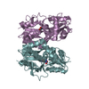
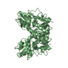
SHEET THE SHEET STRUCTURE OF THIS MOLECULE IS BIFURCATED. IN ORDER TO REPRESENT THIS FEATURE IN ... SHEET THE SHEET STRUCTURE OF THIS MOLECULE IS BIFURCATED. IN ORDER TO REPRESENT THIS FEATURE IN THE SHEET RECORDS BELOW, TWO SHEETS ARE DEFINED.
Mass: 18.015 Da / Num. of mol.: 30 / Source method: isolated from a natural source / Formula: H2O
Compound details
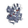
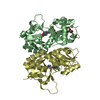
RECEPTOR FOR GLUTAMATE. L-GLUTAMATE ACTS AS AN EXCITATORY NEUROTRANSMITTER AT MANY SYNAPSES IN THE ...RECEPTOR FOR GLUTAMATE. L-GLUTAMATE ACTS AS AN EXCITATORY NEUROTRANSMITTER AT MANY SYNAPSES IN THE CENTRAL NERVOUS SYSTEM. THE POSTSYNAPTIC ACTIONS OF GLU ARE MEDIATED BY A VARIETY OF RECEPTORS THAT ARE NAMED ACCORDING TO THEIR SELECTIVE AGONISTS.
Has protein modification
Y
Sequence details
THE NATIVE GLURDELTA2 IS A MEMBRANE PROTEIN. THE CRYSTALLIZED PROTEIN IS A SE-MET DERIVATIVE OF THE ...THE NATIVE GLURDELTA2 IS A MEMBRANE PROTEIN. THE CRYSTALLIZED PROTEIN IS A SE-MET DERIVATIVE OF THE EXTRACELLULAR LIGAND-BINDING CORE OF GLURDELTA2. TRANSMEMBRANE REGIONS WERE GENETICALLY REMOVED AND REPLACED WITH A GLY-THR LINKER. CONSEQUENTLY, THE PROTEIN SEQUENCE MATCHES DISCONTINOUSLY WITH THE REFERENCE DATABASE. THE FIRST GLYCINE IS A REMNANT OF A TRYPSIN CLEAVAGE SITE.
-
Experimental details
-
Experiment
Experiment
Method: X-RAY DIFFRACTION / Number of used crystals: 1
-
Sample preparation
Crystal
Density Matthews: 2.47 Å3/Da / Density % sol: 50.2 % / Description: NONE
Crystal grow
Method: vapor diffusion, hanging drop / pH: 8.5 Details: 11% PEG4000, 0.1 M TRIS-HCL PH 8.5, 50 MM CACL2. HANGING DROP VAPOR DIFFUSION.
In the structure databanks used in Yorodumi, some data are registered as the other names, "COVID-19 virus" and "2019-nCoV". Here are the details of the virus and the list of structure data.
Jan 31, 2019. EMDB accession codes are about to change! (news from PDBe EMDB page)
EMDB accession codes are about to change! (news from PDBe EMDB page)
The allocation of 4 digits for EMDB accession codes will soon come to an end. Whilst these codes will remain in use, new EMDB accession codes will include an additional digit and will expand incrementally as the available range of codes is exhausted. The current 4-digit format prefixed with “EMD-” (i.e. EMD-XXXX) will advance to a 5-digit format (i.e. EMD-XXXXX), and so on. It is currently estimated that the 4-digit codes will be depleted around Spring 2019, at which point the 5-digit format will come into force.
The EM Navigator/Yorodumi systems omit the EMD- prefix.
Related info.:Q: What is EMD? / ID/Accession-code notation in Yorodumi/EM Navigator
Yorodumi is a browser for structure data from EMDB, PDB, SASBDB, etc.
This page is also the successor to EM Navigator detail page, and also detail information page/front-end page for Omokage search.
The word "yorodu" (or yorozu) is an old Japanese word meaning "ten thousand". "mi" (miru) is to see.
Related info.:EMDB / PDB / SASBDB / Comparison of 3 databanks / Yorodumi Search / Aug 31, 2016. New EM Navigator & Yorodumi / Yorodumi Papers / Jmol/JSmol / Function and homology information / Changes in new EM Navigator and Yorodumi
 Movie
Movie Controller

 Yorodumi
 Open data
 Basic information
 Components
 Keywords
 Function and homology information
 X-RAY DIFFRACTION /
  Authors
 Citation
 Structure visualization
 Downloads & links
 Other downloads


 PDBj
PDBj

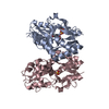
 Assembly
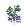


 Mass: 40.078 Da / Num. of mol.: 2 / Source method: obtained synthetically / Formula: Ca
 Mass: 18.015 Da / Num. of mol.: 30 / Source method: isolated from a natural source / Formula: H2O
 Sample preparation
 / Beamline: X12 / Wavelength: 0.9537
  Processing