 Movie
Movie Controller

[English] 日本語
 Yorodumi
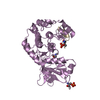
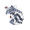
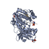
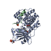
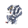
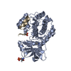
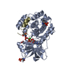
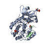
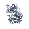
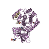
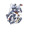
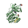
- PDB-2uw4: Structure of PKA-PKB chimera complexed with 2-(4-(5-methyl-1H-pyr... -
+ Open data

- Basic information
| Entry | Database: PDB / ID: 2uw4 | ||||||
|---|---|---|---|---|---|---|---|
| Title | Structure of PKA-PKB chimera complexed with 2-(4-(5-methyl-1H-pyrazol- 4-yl)-phenyl)-ethylamine | ||||||
 Components Components |
| ||||||
 Keywords Keywords | TRANSFERASE / CAMP / KINASE / MYRISTATE / LIPOPROTEIN / SERINE/THREONINE-PROTEIN KINASE / NUCLEOTIDE-BINDING / PROTEIN KINASE INHIBITOR / ATP-BINDING / NUCLEAR PROTEIN / PHOSPHORYLATION | ||||||
| Function / homology |  Function and homology information Function and homology informationCD209 (DC-SIGN) signaling / HDL assembly / Regulation of insulin secretion / Rap1 signalling / Ion homeostasis / negative regulation of cAMP-dependent protein kinase activity / PKA activation in glucagon signalling / DARPP-32 events / CREB1 phosphorylation through the activation of Adenylate Cyclase / GPER1 signaling ...CD209 (DC-SIGN) signaling / HDL assembly / Regulation of insulin secretion / Rap1 signalling / Ion homeostasis / negative regulation of cAMP-dependent protein kinase activity / PKA activation in glucagon signalling / DARPP-32 events / CREB1 phosphorylation through the activation of Adenylate Cyclase / GPER1 signaling / Factors involved in megakaryocyte development and platelet production / Loss of Nlp from mitotic centrosomes / Recruitment of mitotic centrosome proteins and complexes / Loss of proteins required for interphase microtubule organization from the centrosome / Anchoring of the basal body to the plasma membrane / RET signaling / AURKA Activation by TPX2 / Interleukin-3, Interleukin-5 and GM-CSF signaling / Recruitment of NuMA to mitotic centrosomes / VEGFA-VEGFR2 Pathway / PKA activation / MAPK6/MAPK4 signaling / GLI3 is processed to GLI3R by the proteasome / Regulation of PLK1 Activity at G2/M Transition / Hedgehog 'off' state / cAMP-dependent protein kinase inhibitor activity / histone H1-4S35 kinase activity / cAMP-dependent protein kinase / regulation of protein processing / cAMP-dependent protein kinase activity / cellular response to parathyroid hormone stimulus / protein localization to lipid droplet / regulation of bicellular tight junction assembly / cAMP-dependent protein kinase complex / negative regulation of protein import into nucleus / dorsal/ventral neural tube patterning / interleukin-2-mediated signaling pathway / Mitochondrial protein degradation / regulation of osteoblast differentiation / Glucagon-like Peptide-1 (GLP1) regulates insulin secretion / sperm capacitation / cellular response to cold / High laminar flow shear stress activates signaling by PIEZO1 and PECAM1:CDH5:KDR in endothelial cells / Vasopressin regulates renal water homeostasis via Aquaporins / protein kinase A regulatory subunit binding / negative regulation of glycolytic process through fructose-6-phosphate / ciliary base / protein kinase A catalytic subunit binding / intracellular potassium ion homeostasis / mesoderm formation / TORC1 signaling / plasma membrane raft / axoneme / sperm flagellum / postsynaptic modulation of chemical synaptic transmission / negative regulation of cAMP/PKA signal transduction / regulation of G2/M transition of mitotic cell cycle / negative regulation of TORC1 signaling / protein serine/threonine/tyrosine kinase activity / regulation of proteasomal protein catabolic process / protein export from nucleus / cellular response to glucagon stimulus / cellular response to nutrient levels / positive regulation of phagocytosis / positive regulation of gluconeogenesis / acrosomal vesicle / positive regulation of protein export from nucleus / neuromuscular junction / negative regulation of smoothened signaling pathway / neural tube closure / cellular response to glucose stimulus / positive regulation of cholesterol biosynthetic process / mRNA processing / adenylate cyclase-inhibiting G protein-coupled receptor signaling pathway / positive regulation of insulin secretion / manganese ion binding / cellular response to heat / adenylate cyclase-activating G protein-coupled receptor signaling pathway / protein kinase activity / regulation of cell cycle / nuclear speck / postsynapse / protein domain specific binding / protein serine kinase activity / protein serine/threonine kinase activity / ubiquitin protein ligase binding / centrosome / protein kinase binding / perinuclear region of cytoplasm / glutamatergic synapse / magnesium ion binding / negative regulation of transcription by RNA polymerase II / mitochondrion / ATP binding / nucleus / cytoplasm / cytosol Similarity search - Function | ||||||
| Biological species |   HOMO SAPIENS (human) HOMO SAPIENS (human) | ||||||
| Method |  X-RAY DIFFRACTION / MOLECULAR REPLACEMENT / Resolution: 2 Å X-RAY DIFFRACTION / MOLECULAR REPLACEMENT / Resolution: 2 Å | ||||||
 Authors Authors | Davies, T.G. / Saxty, G. / Woodhead, S.J. / Berdini, V. / Verdonk, M.L. / Wyatt, P.G. / Boyle, R.G. / Barford, D. / Downham, R. / Garrett, M.D. / Carr, R.A. | ||||||
 Citation Citation | Journal: J.Med.Chem. / Year: 2007 Title: Identification of Inhibitors of Protein Kinase B Using Fragment-Based Lead Discovery Authors: Saxty, G. / Woodhead, S.J. / Berdini, V. / Davies, T.G. / Verdonk, M.L. / Wyatt, P.G. / Boyle, R.G. / Barford, D. / Downham, R. / Garrett, M.D. / Carr, R.A. | ||||||
| History |
|
- Structure visualization
| Structure viewer | Molecule: MolmilJmol/JSmol |
|---|
- Downloads & links
-Download
| PDBx/mmCIF format | 2uw4.cif.gz | 96.7 KB | Display | PDBx/mmCIF format |
|---|---|---|---|---|
| PDB format | pdb2uw4.ent.gz | 73 KB | Display | PDB format |
| PDBx/mmJSON format | 2uw4.json.gz | Tree view | PDBx/mmJSON format | |
| Others |  Other downloads Other downloads |
-Validation report
| Arichive directory | https://data.pdbj.org/pub/pdb/validation_reports/uw/2uw4ftp://data.pdbj.org/pub/pdb/validation_reports/uw/2uw4 | HTTPS FTP |
|---|
-Related structure data
| Related structure data |  2uw3C  2uw5C  2uw6C  2uw7C  2uw8C  2uw9C C: citing same article ( |
|---|---|
| Similar structure data |


-Links
 PDBj
PDBj


- Assembly
| Deposited unit | 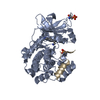
| ||||||||
|---|---|---|---|---|---|---|---|---|---|
| 1 |
| ||||||||
| Unit cell |
|
-Components
| #1: Protein | Mass: 40830.617 Da / Num. of mol.: 1 / Mutation: YES Source method: isolated from a genetically manipulated source Source: (gene. exp.)  |
|---|---|
| #2: Protein/peptide | Mass: 2226.411 Da / Num. of mol.: 1 / Fragment: RESIDUES 5-24 / Source method: obtained synthetically / Source: (synth.) HOMO SAPIENS (human) / References: UniProt: P61925 |
| #3: Chemical | ChemComp-L15 /   Mass: 201.268 Da / Num. of mol.: 1 / Source method: obtained synthetically / Formula: C12H15N3 Mass: 201.268 Da / Num. of mol.: 1 / Source method: obtained synthetically / Formula: C12H15N3 |
| #4: Water | ChemComp-HOH /  Mass: 18.015 Da / Num. of mol.: 391 / Source method: isolated from a natural source / Formula: H2O Mass: 18.015 Da / Num. of mol.: 391 / Source method: isolated from a natural source / Formula: H2O |
| Compound details | ENGINEERED RESIDUE IN CHAIN A, VAL 104 TO THR ENGINEERED RESIDUE IN CHAIN A, VAL 123 TO ALA ...ENGINEERED |
| Has protein modification | Y |
| Sequence details | 4 RESIDUES OF THE WILD-TYPE PKA SEQUENCE HAVE BEEN MUTATED TO THE CORRESPOND |
-Experimental details
-Experiment
| Experiment | Method: X-RAY DIFFRACTION |
|---|
- Sample preparation
| Crystal | Density Matthews: 2.32 Å3/Da / Density % sol: 46.5 % |
|---|
-Data collection
| Diffraction | Mean temperature: 100 K |
|---|---|
| Diffraction source | Source: ROTATING ANODE / Wavelength: 1.5418 |
| Detector | Type: RIGAKU IMAGE PLATE / Detector: IMAGE PLATE |
| Radiation | Protocol: SINGLE WAVELENGTH / Monochromatic (M) / Laue (L): M / Scattering type: x-ray |
| Radiation wavelength | Wavelength: 1.5418 Å / Relative weight: 1 |
| Reflection | Resolution: 2→33.3 Å / Num. obs: 28339 / % possible obs: 97.1 % / Redundancy: 2.9 % / Rmerge(I) obs: 0.12 / Net I/σ(I): 4.2 |
| Reflection shell | Resolution: 2→2.07 Å / Redundancy: 2.5 % / Rmerge(I) obs: 0.36 / Mean I/σ(I) obs: 1.7 / % possible all: 91.4 |
- Processing
| Software | Name: REFMAC / Version: 5.2.0019G / Classification: refinement | ||||||||||||||||||||||||||||||||||||||||||||||||||||||||||||||||||||||||||||||||||||||||||||||||||||||||||||||||||||||||||||||||||||||||||||||||||||||||||||||||||||||||||||||||||||||
|---|---|---|---|---|---|---|---|---|---|---|---|---|---|---|---|---|---|---|---|---|---|---|---|---|---|---|---|---|---|---|---|---|---|---|---|---|---|---|---|---|---|---|---|---|---|---|---|---|---|---|---|---|---|---|---|---|---|---|---|---|---|---|---|---|---|---|---|---|---|---|---|---|---|---|---|---|---|---|---|---|---|---|---|---|---|---|---|---|---|---|---|---|---|---|---|---|---|---|---|---|---|---|---|---|---|---|---|---|---|---|---|---|---|---|---|---|---|---|---|---|---|---|---|---|---|---|---|---|---|---|---|---|---|---|---|---|---|---|---|---|---|---|---|---|---|---|---|---|---|---|---|---|---|---|---|---|---|---|---|---|---|---|---|---|---|---|---|---|---|---|---|---|---|---|---|---|---|---|---|---|---|---|---|
| Refinement | Method to determine structure: MOLECULAR REPLACEMENT / Resolution: 2→33.32 Å / Cor.coef. Fo:Fc: 0.936 / Cor.coef. Fo:Fc free: 0.901 / SU B: 6.046 / SU ML: 0.164 / Cross valid method: THROUGHOUT / ESU R: 0.207 / ESU R Free: 0.192 / Stereochemistry target values: MAXIMUM LIKELIHOOD
| ||||||||||||||||||||||||||||||||||||||||||||||||||||||||||||||||||||||||||||||||||||||||||||||||||||||||||||||||||||||||||||||||||||||||||||||||||||||||||||||||||||||||||||||||||||||
| Solvent computation | Ion probe radii: 0.8 Å / Shrinkage radii: 0.8 Å / VDW probe radii: 1.2 Å / Solvent model: BABINET MODEL WITH MASK | ||||||||||||||||||||||||||||||||||||||||||||||||||||||||||||||||||||||||||||||||||||||||||||||||||||||||||||||||||||||||||||||||||||||||||||||||||||||||||||||||||||||||||||||||||||||
| Displacement parameters | Biso mean: 21.69 Å2
| ||||||||||||||||||||||||||||||||||||||||||||||||||||||||||||||||||||||||||||||||||||||||||||||||||||||||||||||||||||||||||||||||||||||||||||||||||||||||||||||||||||||||||||||||||||||
| Refinement step | Cycle: LAST / Resolution: 2→33.32 Å
| ||||||||||||||||||||||||||||||||||||||||||||||||||||||||||||||||||||||||||||||||||||||||||||||||||||||||||||||||||||||||||||||||||||||||||||||||||||||||||||||||||||||||||||||||||||||
| Refine LS restraints |
|