 Movie
Movie Controller


+ Open data

- Basic information
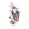
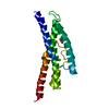
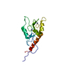
| Entry | Database: PDB / ID: 2uv1 | ||||||
|---|---|---|---|---|---|---|---|
| Title | Hexagonal crystal form of GamS from bacteriophage lambda. | ||||||
 Components Components | HOST-NUCLEASE INHIBITOR PROTEIN GAM | ||||||
 Keywords Keywords | INHIBITOR / PUTATIVE DNA MIMETIC / NUCLEASE INHIBITOR RECBCD INHIBITOR / BACTERIOPHAGE LAMBDA | ||||||
| Function / homology |  Function and homology information Function and homology informationsymbiont-mediated evasion of DNA end degradation by host / deoxyribonuclease inhibitor activity / symbiont-mediated suppression of host innate immune response Similarity search - Function | ||||||
| Biological species |  BACTERIOPHAGE LAMBDA (virus) BACTERIOPHAGE LAMBDA (virus) | ||||||
| Method |  X-RAY DIFFRACTION / SYNCHROTRON / MOLECULAR REPLACEMENT / Resolution: 2.6 Å X-RAY DIFFRACTION / SYNCHROTRON / MOLECULAR REPLACEMENT / Resolution: 2.6 Å | ||||||
 Authors Authors | Court, R.I. / Cook, N. / Saikrishnan, K. / Wigley, D.B. | ||||||
 Citation Citation | Journal: J.Mol.Biol. / Year: 2007 Title: The Crystal Structure of Lambda-Gam Protein Suggests a Model for Recbcd Inhibition. Authors: Court, R.I. / Cook, N. / Saikrishnan, K. / Wigley, D.B. | ||||||
| History |
|
- Structure visualization
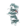
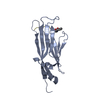
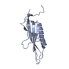
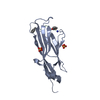
| Structure viewer | Molecule: MolmilJmol/JSmol |
|---|
- Downloads & links
-Download
| PDBx/mmCIF format | 2uv1.cif.gz | 44.5 KB | Display | PDBx/mmCIF format |
|---|---|---|---|---|
| PDB format | pdb2uv1.ent.gz | 31.5 KB | Display | PDB format |
| PDBx/mmJSON format | 2uv1.json.gz | Tree view | PDBx/mmJSON format | |
| Others |  Other downloads Other downloads |
-Validation report
| Summary document | 2uv1_validation.pdf.gz | 434.6 KB | Display | wwPDB validaton report |
|---|---|---|---|---|
| Full document | 2uv1_full_validation.pdf.gz | 438 KB | Display | |
| Data in XML | 2uv1_validation.xml.gz | 8.2 KB | Display | |
| Data in CIF | 2uv1_validation.cif.gz | 10.5 KB | Display | |
| Arichive directory | https://data.pdbj.org/pub/pdb/validation_reports/uv/2uv1ftp://data.pdbj.org/pub/pdb/validation_reports/uv/2uv1 | HTTPS FTP |
-Related structure data
| Related structure data |  2uuzSC S: Starting model for refinement C: citing same article ( |
|---|---|
| Similar structure data |


-Links
 PDBj
PDBj- Assembly
| Deposited unit | 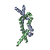
| ||||||||
|---|---|---|---|---|---|---|---|---|---|
| 1 |
| ||||||||
| Unit cell |
|
-Components
| #1: Protein | Mass: 11733.002 Da / Num. of mol.: 2 / Fragment: RESIDUES 40-138 Source method: isolated from a genetically manipulated source Source: (gene. exp.) BACTERIOPHAGE LAMBDA (virus) / Plasmid: PKM574 / Production host:  #2: Water | ChemComp-HOH / |  Mass: 18.015 Da / Num. of mol.: 31 / Source method: isolated from a natural source / Formula: H2O Mass: 18.015 Da / Num. of mol.: 31 / Source method: isolated from a natural source / Formula: H2OSequence details | CONSTRUCT STARTS AT RESIDUE 40 OF REPORTED SEQUENCE. | |
|---|
-Experimental details
-Experiment
| Experiment | Method: X-RAY DIFFRACTION / Number of used crystals: 1 |
|---|
- Sample preparation
| Crystal | Density Matthews: 2.93 Å3/Da / Density % sol: 41.5 % |
|---|---|
| Crystal grow | pH: 6.5 / Details: 100 MM MES, PH 6.5, 20-25 % PEG 400, 0.1 M NACL |
-Data collection
| Diffraction | Mean temperature: 100 K |
|---|---|
| Diffraction source | Source: SYNCHROTRON / Site: ESRF  / Beamline: ID14-4 / Wavelength: 0.9794 / Beamline: ID14-4 / Wavelength: 0.9794 |
| Detector | Type: ADSC CCD / Detector: CCD |
| Radiation | Protocol: SINGLE WAVELENGTH / Monochromatic (M) / Laue (L): M / Scattering type: x-ray |
| Radiation wavelength | Wavelength: 0.9794 Å / Relative weight: 1 |
| Reflection | Resolution: 2.6→73 Å / Num. obs: 6851 / % possible obs: 99.5 % / Observed criterion σ(I): 2 / Redundancy: 15.3 % / Rmerge(I) obs: 0.09 / Net I/σ(I): 6 |
| Reflection shell | Resolution: 2.6→2.74 Å / Redundancy: 16.2 % / Rmerge(I) obs: 0.27 / Mean I/σ(I) obs: 2.4 / % possible all: 99.5 |
- Processing
| Software |
| ||||||||||||||||||||||||||||||||||||||||||||||||||||||||||||||||||||||||||||||||||||||||||||||||||||||||||||||||||||||||||||||||||||||||||||||||||||||||||||||||||||||||||||||||||||||
|---|---|---|---|---|---|---|---|---|---|---|---|---|---|---|---|---|---|---|---|---|---|---|---|---|---|---|---|---|---|---|---|---|---|---|---|---|---|---|---|---|---|---|---|---|---|---|---|---|---|---|---|---|---|---|---|---|---|---|---|---|---|---|---|---|---|---|---|---|---|---|---|---|---|---|---|---|---|---|---|---|---|---|---|---|---|---|---|---|---|---|---|---|---|---|---|---|---|---|---|---|---|---|---|---|---|---|---|---|---|---|---|---|---|---|---|---|---|---|---|---|---|---|---|---|---|---|---|---|---|---|---|---|---|---|---|---|---|---|---|---|---|---|---|---|---|---|---|---|---|---|---|---|---|---|---|---|---|---|---|---|---|---|---|---|---|---|---|---|---|---|---|---|---|---|---|---|---|---|---|---|---|---|---|
| Refinement | Method to determine structure: MOLECULAR REPLACEMENT Starting model: PDB ENTRY 2UUZ Resolution: 2.6→105.41 Å / Cor.coef. Fo:Fc: 0.906 / Cor.coef. Fo:Fc free: 0.838 / SU B: 11.503 / SU ML: 0.258 / Cross valid method: THROUGHOUT / ESU R: 0.564 / ESU R Free: 0.348 / Stereochemistry target values: MAXIMUM LIKELIHOOD Details: HYDROGENS HAVE BEEN ADDED IN THE RIDING POSITIONS. N-TERMINAL RESIDUES 40-59 OF CHAIN A AND 40- 58 OF CHAIN B ARE DISORDERED. THE C-TERMINAL RESIDUE, VAL 138, IS DISORDERED IN BOTH CHAINS.
| ||||||||||||||||||||||||||||||||||||||||||||||||||||||||||||||||||||||||||||||||||||||||||||||||||||||||||||||||||||||||||||||||||||||||||||||||||||||||||||||||||||||||||||||||||||||
| Solvent computation | Ion probe radii: 0.8 Å / Shrinkage radii: 0.8 Å / VDW probe radii: 1.4 Å / Solvent model: MASK | ||||||||||||||||||||||||||||||||||||||||||||||||||||||||||||||||||||||||||||||||||||||||||||||||||||||||||||||||||||||||||||||||||||||||||||||||||||||||||||||||||||||||||||||||||||||
| Displacement parameters | Biso mean: 35.47 Å2
| ||||||||||||||||||||||||||||||||||||||||||||||||||||||||||||||||||||||||||||||||||||||||||||||||||||||||||||||||||||||||||||||||||||||||||||||||||||||||||||||||||||||||||||||||||||||
| Refinement step | Cycle: LAST / Resolution: 2.6→105.41 Å
| ||||||||||||||||||||||||||||||||||||||||||||||||||||||||||||||||||||||||||||||||||||||||||||||||||||||||||||||||||||||||||||||||||||||||||||||||||||||||||||||||||||||||||||||||||||||
| Refine LS restraints |
|