 Movie
Movie Controller

[English] 日本語
 Yorodumi
- PDB-2uup: Crystal structure of MurD ligase in complex with D-Glu containing... -
+ Open data

- Basic information
| Entry | Database: PDB / ID: 2uup | ||||||
|---|---|---|---|---|---|---|---|
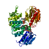
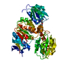
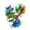
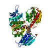
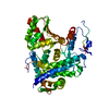
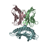
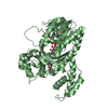
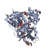
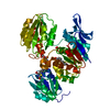
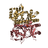
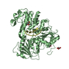
| Title | Crystal structure of MurD ligase in complex with D-Glu containing sulfonamide inhibitor | ||||||
 Components Components | UDP-N-ACETYLMURAMOYLALANINE--D-GLUTAMATE LIGASE | ||||||
 Keywords Keywords | LIGASE / MURD-INHIBITOR COMPLEX / PEPTIDOGLYCAN SYNTHESIS / CELL WALL / CELL SHAPE / CELL CYCLE / NUCLEOTIDE-BINDING / SULFONAMIDE INHIBITOR / MURD LIGASE / ATP-BINDING / CELL DIVISION | ||||||
| Function / homology |  Function and homology information Function and homology informationUDP-N-acetylmuramoyl-L-alanine-D-glutamate ligase / UDP-N-acetylmuramoylalanine-D-glutamate ligase activity / peptidoglycan biosynthetic process / cell wall organization / regulation of cell shape / cell division / ATP binding / identical protein binding / cytoplasm Similarity search - Function | ||||||
| Biological species |  | ||||||
| Method |  X-RAY DIFFRACTION / SYNCHROTRON / MOLECULAR REPLACEMENT / Resolution: 1.88 Å X-RAY DIFFRACTION / SYNCHROTRON / MOLECULAR REPLACEMENT / Resolution: 1.88 Å | ||||||
 Authors Authors | Humljan, J. / Kotnik, M. / Contreras-Martel, C. / Blanot, D. / Urleb, U. / Dessen, A. / Solmajer, T. / Gobec, S. | ||||||
 Citation Citation | Journal: J. Med. Chem. / Year: 2008 Title: Novel naphthalene-N-sulfonyl-D-glutamic acid derivatives as inhibitors of MurD, a key peptidoglycan biosynthesis enzyme. Authors: Humljan, J. / Kotnik, M. / Contreras-Martel, C. / Blanot, D. / Urleb, U. / Dessen, A. / Solmajer, T. / Gobec, S. | ||||||
| History |
|
- Structure visualization
| Structure viewer | Molecule: MolmilJmol/JSmol |
|---|
- Downloads & links
-Download
| PDBx/mmCIF format | 2uup.cif.gz | 108.1 KB | Display | PDBx/mmCIF format |
|---|---|---|---|---|
| PDB format | pdb2uup.ent.gz | 80.6 KB | Display | PDB format |
| PDBx/mmJSON format | 2uup.json.gz | Tree view | PDBx/mmJSON format | |
| Others |  Other downloads Other downloads |
-Validation report
| Arichive directory | https://data.pdbj.org/pub/pdb/validation_reports/uu/2uupftp://data.pdbj.org/pub/pdb/validation_reports/uu/2uup | HTTPS FTP |
|---|
-Related structure data
| Related structure data |  2uuoC  2vtdC  2vteC  3uagS S: Starting model for refinement C: citing same article ( |
|---|---|
| Similar structure data |


-Links
 PDBj
PDBj- Assembly
| Deposited unit | 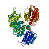
| ||||||||
|---|---|---|---|---|---|---|---|---|---|
| 1 |
| ||||||||
| Unit cell |
|
-Components
| #1: Protein | Mass: 47979.398 Da / Num. of mol.: 1 Source method: isolated from a genetically manipulated source Details: INHIBITOR N-(6-(4-CYANO-BENZYLOXY)-NAPHTHALENE-2-SULFONYL)-D-GLUTAMIC ACID BOUND Source: (gene. exp.) References: UniProt: P14900, UDP-N-acetylmuramoyl-L-alanine-D-glutamate ligase | ||||
|---|---|---|---|---|---|
| #2: Chemical | ChemComp-LK4 /   Mass: 468.479 Da / Num. of mol.: 1 / Source method: obtained synthetically / Formula: C23H20N2O7S Mass: 468.479 Da / Num. of mol.: 1 / Source method: obtained synthetically / Formula: C23H20N2O7S | ||||
| #3: Chemical |   Mass: 96.063 Da / Num. of mol.: 3 / Source method: obtained synthetically / Formula: SO4 Mass: 96.063 Da / Num. of mol.: 3 / Source method: obtained synthetically / Formula: SO4#4: Water | ChemComp-HOH / |  Mass: 18.015 Da / Num. of mol.: 418 / Source method: isolated from a natural source / Formula: H2O Mass: 18.015 Da / Num. of mol.: 418 / Source method: isolated from a natural source / Formula: H2OSequence details | C-TERMINAL HIS-TAG (SHHHHHH) | |
-Experimental details
-Experiment
| Experiment | Method: X-RAY DIFFRACTION / Number of used crystals: 1 |
|---|
- Sample preparation
| Crystal | Density Matthews: 3.1 Å3/Da / Density % sol: 60 % / Description: NONE |
|---|---|
| Crystal grow | pH: 7.5 Details: PROTEIN WAS CRYSTALLIZED FROM 1.7 M (NH4)2SO4, 7% PEG 400, 100 MM HEPES, PH 7.5; THEN SOAKED IN 1 MM INHIBITOR SOLUTION. |
-Data collection
| Diffraction | Mean temperature: 288 K |
|---|---|
| Diffraction source | Source: SYNCHROTRON / Site: ESRF  / Beamline: BM30A / Wavelength: 0.979 / Beamline: BM30A / Wavelength: 0.979 |
| Detector | Type: MARRESEARCH / Detector: CCD / Date: Jun 15, 2006 |
| Radiation | Protocol: SINGLE WAVELENGTH / Monochromatic (M) / Laue (L): M / Scattering type: x-ray |
| Radiation wavelength | Wavelength: 0.979 Å / Relative weight: 1 |
| Reflection | Resolution: 1.88→50 Å / Num. obs: 45633 / % possible obs: 98.1 % / Observed criterion σ(I): 3 / Redundancy: 5.1 % / Rmerge(I) obs: 0.06 / Net I/σ(I): 19.6 |
| Reflection shell | Resolution: 1.88→1.99 Å / Redundancy: 5.1 % / Rmerge(I) obs: 0.28 / Mean I/σ(I) obs: 6.1 / % possible all: 95.1 |
- Processing
| Software |
| ||||||||||||||||||||||||||||||||||||||||||||||||||||||||||||||||||||||||||||||||||||||||||||||||||||||||||||||||||||||||||||||||||||||||||||||||||||||||||||||||||||||||||||||||||||||
|---|---|---|---|---|---|---|---|---|---|---|---|---|---|---|---|---|---|---|---|---|---|---|---|---|---|---|---|---|---|---|---|---|---|---|---|---|---|---|---|---|---|---|---|---|---|---|---|---|---|---|---|---|---|---|---|---|---|---|---|---|---|---|---|---|---|---|---|---|---|---|---|---|---|---|---|---|---|---|---|---|---|---|---|---|---|---|---|---|---|---|---|---|---|---|---|---|---|---|---|---|---|---|---|---|---|---|---|---|---|---|---|---|---|---|---|---|---|---|---|---|---|---|---|---|---|---|---|---|---|---|---|---|---|---|---|---|---|---|---|---|---|---|---|---|---|---|---|---|---|---|---|---|---|---|---|---|---|---|---|---|---|---|---|---|---|---|---|---|---|---|---|---|---|---|---|---|---|---|---|---|---|---|---|
| Refinement | Method to determine structure: MOLECULAR REPLACEMENT Starting model: PDB ENTRY 3UAG Resolution: 1.88→65.51 Å / Cor.coef. Fo:Fc: 0.944 / Cor.coef. Fo:Fc free: 0.914 / Cross valid method: THROUGHOUT / ESU R: 0.127 / ESU R Free: 0.125 / Stereochemistry target values: MAXIMUM LIKELIHOOD
| ||||||||||||||||||||||||||||||||||||||||||||||||||||||||||||||||||||||||||||||||||||||||||||||||||||||||||||||||||||||||||||||||||||||||||||||||||||||||||||||||||||||||||||||||||||||
| Solvent computation | Ion probe radii: 0.8 Å / Shrinkage radii: 0.8 Å / VDW probe radii: 1.4 Å / Solvent model: MASK | ||||||||||||||||||||||||||||||||||||||||||||||||||||||||||||||||||||||||||||||||||||||||||||||||||||||||||||||||||||||||||||||||||||||||||||||||||||||||||||||||||||||||||||||||||||||
| Displacement parameters | Biso mean: 19.82 Å2
| ||||||||||||||||||||||||||||||||||||||||||||||||||||||||||||||||||||||||||||||||||||||||||||||||||||||||||||||||||||||||||||||||||||||||||||||||||||||||||||||||||||||||||||||||||||||
| Refinement step | Cycle: LAST / Resolution: 1.88→65.51 Å
| ||||||||||||||||||||||||||||||||||||||||||||||||||||||||||||||||||||||||||||||||||||||||||||||||||||||||||||||||||||||||||||||||||||||||||||||||||||||||||||||||||||||||||||||||||||||
| Refine LS restraints |
|