- PDB-2rko: Crystal Structure of the Vps4p-dimer -
+
Open data
ID or keywords:
Loading...
-
Basic information
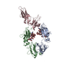
Entry
Database: PDB / ID: 2rko
Title
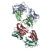
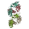
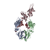
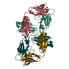
Crystal Structure of the Vps4p-dimer
Components
Vacuolar protein sorting-associated protein 4
Keywords
PROTEIN TRANSPORT / AAA-ATPase domain / ATP-binding / Endosome / Membrane / Nucleotide-binding / Phosphorylation / Transport
Function / homology
Function and homology information
ESCRT IV complex / Sealing of the nuclear envelope (NE) by ESCRT-III / late endosome to lysosome transport via multivesicular body sorting pathway / intralumenal vesicle formation / : / protein retention in Golgi apparatus / Endosomal Sorting Complex Required For Transport (ESCRT) / late endosome to vacuole transport via multivesicular body sorting pathway / sterol metabolic process / nuclear membrane reassembly ...ESCRT IV complex / Sealing of the nuclear envelope (NE) by ESCRT-III / late endosome to lysosome transport via multivesicular body sorting pathway / intralumenal vesicle formation / : / protein retention in Golgi apparatus / Endosomal Sorting Complex Required For Transport (ESCRT) / late endosome to vacuole transport via multivesicular body sorting pathway / sterol metabolic process / nuclear membrane reassembly / multivesicular body sorting pathway / vacuole organization / midbody abscission / membrane fission / late endosome to vacuole transport / multivesicular body assembly / ubiquitin-dependent protein catabolic process via the multivesicular body sorting pathway / plasma membrane repair / reticulophagy / endosomal transport / ATPase complex / nucleus organization / autophagosome maturation / nuclear pore / macroautophagy / autophagy / protein transport / midbody / endosome / endoplasmic reticulum / protein homodimerization activity / ATP hydrolysis activity / ATP binding / membrane / identical protein binding / plasma membrane / cytoplasm Similarity search - Function
Vacuolar protein sorting-associated protein 4, MIT domain / MIT (microtubule interacting and transport) domain / MIT domain superfamily / Vps4 oligomerisation, C-terminal / MIT domain / Microtubule Interacting and Trafficking molecule domain / : / Vps4 C terminal oligomerisation domain / Helicase, Ruva Protein; domain 3 - #60 / Helicase, Ruva Protein; domain 3 ...Vacuolar protein sorting-associated protein 4, MIT domain / MIT (microtubule interacting and transport) domain / MIT domain superfamily / Vps4 oligomerisation, C-terminal / MIT domain / Microtubule Interacting and Trafficking molecule domain / : / Vps4 C terminal oligomerisation domain / Helicase, Ruva Protein; domain 3 - #60 / Helicase, Ruva Protein; domain 3 / AAA ATPase, AAA+ lid domain / AAA+ lid domain / ATPase, AAA-type, conserved site / AAA-protein family signature. / ATPase family associated with various cellular activities (AAA) / ATPase, AAA-type, core / P-loop containing nucleotide triphosphate hydrolases / ATPases associated with a variety of cellular activities / AAA+ ATPase domain / Rossmann fold / P-loop containing nucleoside triphosphate hydrolase / Orthogonal Bundle / 3-Layer(aba) Sandwich / Mainly Alpha / Alpha Beta Similarity search - Domain/homology
Journal: J Mol Biol / Year: 2008 Title: Vacuolar protein sorting: two different functional states of the AAA-ATPase Vps4p. Authors: Claudia Hartmann / Mohamed Chami / Ulrich Zachariae / Bert L de Groot / Andreas Engel / Markus G Grütter / Abstract: The vacuolar protein sorting (Vps) pathway, in which Vps4 class I AAA-ATPases play a central role, regulates growth factor receptors, immune response, and developmental signaling, and participates in ...The vacuolar protein sorting (Vps) pathway, in which Vps4 class I AAA-ATPases play a central role, regulates growth factor receptors, immune response, and developmental signaling, and participates in tumor suppression, apoptosis, and retrovirus budding. We present the first atomic structure of the nucleotide-free yeast His(6)DeltaNVps4p dimer and its AMPPNP (5'-adenylyl-beta,gamma-imidodiphosphate)-bound tetradecamer, derived from a cryo electron microscopy map. Vps4p dimers form two distinct heptameric rings and accommodate AAA cassettes in a head-to-head--not in a head-to-tail-fashion as in class II AAA-ATPases. Our model suggests a mechanism for disassembling ESCRT (endosomal sorting complex required for transport) complexes by movements of substrate-binding domains located at the periphery of the tetradecamer during ATP hydrolysis in one ring, followed by translocation through the central pore and ATP hydrolysis in the second ring.
In the structure databanks used in Yorodumi, some data are registered as the other names, "COVID-19 virus" and "2019-nCoV". Here are the details of the virus and the list of structure data.
Jan 31, 2019. EMDB accession codes are about to change! (news from PDBe EMDB page)
EMDB accession codes are about to change! (news from PDBe EMDB page)
The allocation of 4 digits for EMDB accession codes will soon come to an end. Whilst these codes will remain in use, new EMDB accession codes will include an additional digit and will expand incrementally as the available range of codes is exhausted. The current 4-digit format prefixed with “EMD-” (i.e. EMD-XXXX) will advance to a 5-digit format (i.e. EMD-XXXXX), and so on. It is currently estimated that the 4-digit codes will be depleted around Spring 2019, at which point the 5-digit format will come into force.
The EM Navigator/Yorodumi systems omit the EMD- prefix.
Related info.:Q: What is EMD? / ID/Accession-code notation in Yorodumi/EM Navigator
Yorodumi is a browser for structure data from EMDB, PDB, SASBDB, etc.
This page is also the successor to EM Navigator detail page, and also detail information page/front-end page for Omokage search.
The word "yorodu" (or yorozu) is an old Japanese word meaning "ten thousand". "mi" (miru) is to see.
Related info.:EMDB / PDB / SASBDB / Comparison of 3 databanks / Yorodumi Search / Aug 31, 2016. New EM Navigator & Yorodumi / Yorodumi Papers / Jmol/JSmol / Function and homology information / Changes in new EM Navigator and Yorodumi
 Movie
Movie Controller


 Open data
 Basic information
 Components
 Keywords
 Function and homology information
 X-RAY DIFFRACTION /
  Authors
 Citation
 Structure visualization
 Downloads & links
 Other downloads


 PDBj
PDBj


 Assembly


 Sample preparation
 Processing