 Movie
Movie Controller

[English] 日本語
 Yorodumi
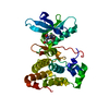
- PDB-2rf9: Crystal structure of the complex between the EGFR kinase domain a... -
+ Open data

- Basic information
| Entry | Database: PDB / ID: 2rf9 | ||||||
|---|---|---|---|---|---|---|---|
| Title | Crystal structure of the complex between the EGFR kinase domain and a Mig6 peptide | ||||||
 Components Components |
| ||||||
 Keywords Keywords | TRANSFERASE / kinase domain / inhibition / dimer / Alternative splicing / Anti-oncogene / ATP-binding / Cell cycle / Disease mutation / Glycoprotein / Membrane / Nucleotide-binding / Phosphorylation / Polymorphism / Receptor / Secreted / Transmembrane / Tyrosine-protein kinase / Ubl conjugation / Cytoplasm | ||||||
| Function / homology |  Function and homology information Function and homology informationregulation of type B pancreatic cell proliferation / negative regulation of epidermal growth factor-activated receptor activity / lung epithelium development / lung vasculature development / negative regulation of protein autophosphorylation / regulation of keratinocyte differentiation / skin morphogenesis / negative regulation of collagen biosynthetic process / lung alveolus development / cellular hyperosmotic response ...regulation of type B pancreatic cell proliferation / negative regulation of epidermal growth factor-activated receptor activity / lung epithelium development / lung vasculature development / negative regulation of protein autophosphorylation / regulation of keratinocyte differentiation / skin morphogenesis / negative regulation of collagen biosynthetic process / lung alveolus development / cellular hyperosmotic response / multivesicular body, internal vesicle lumen / negative regulation of cardiocyte differentiation / Shc-EGFR complex / positive regulation of protein kinase C signaling / Inhibition of Signaling by Overexpressed EGFR / EGFR interacts with phospholipase C-gamma / epidermal growth factor receptor activity / cellular response to dexamethasone stimulus / epidermal growth factor binding / response to UV-A / regulation of peptidyl-tyrosine phosphorylation / negative regulation of cardiac muscle hypertrophy in response to stress / PLCG1 events in ERBB2 signaling / negative regulation of interleukin-1 beta production / ERBB2-EGFR signaling pathway / ERBB2 Activates PTK6 Signaling / PTK6 promotes HIF1A stabilization / Signaling by EGFR / positive regulation of toll-like receptor 9 signaling pathway / intracellular vesicle / negative regulation of epidermal growth factor receptor signaling pathway / ERBB2 Regulates Cell Motility / Developmental Lineage of Mammary Gland Myoepithelial Cells / protein insertion into membrane / Respiratory syncytial virus (RSV) attachment and entry / Signaling by ERBB4 / PI3K events in ERBB2 signaling / positive regulation of cGAS/STING signaling pathway / ossification / positive regulation of phosphorylation / negative regulation of tumor necrosis factor production / positive regulation of peptidyl-serine phosphorylation / Estrogen-dependent nuclear events downstream of ESR-membrane signaling / MAP kinase kinase kinase activity / GAB1 signalosome / protein tyrosine kinase activator activity / negative regulation of macroautophagy / positive regulation of G1/S transition of mitotic cell cycle / Signaling by ERBB2 / TFAP2 (AP-2) family regulates transcription of growth factors and their receptors / transmembrane receptor protein tyrosine kinase activity / positive regulation of epithelial cell proliferation / cellular response to platelet-derived growth factor stimulus / GRB2 events in EGFR signaling / SHC1 events in EGFR signaling / EGFR Transactivation by Gastrin / GTPase activator activity / GRB2 events in ERBB2 signaling / positive regulation of fibroblast proliferation / cellular response to epidermal growth factor stimulus / basal plasma membrane / SHC1 events in ERBB2 signaling / positive regulation of DNA replication / positive regulation of DNA repair / cellular response to amino acid stimulus / phosphatidylinositol 3-kinase/protein kinase B signal transduction / Signal transduction by L1 / cellular response to estradiol stimulus / SH3 domain binding / negative regulation of protein catabolic process / positive regulation of protein localization to plasma membrane / clathrin-coated endocytic vesicle membrane / cell-cell adhesion / NOTCH3 Activation and Transmission of Signal to the Nucleus / Signaling by ERBB2 TMD/JMD mutants / negative regulation of ERK1 and ERK2 cascade / EGFR downregulation / receptor protein-tyrosine kinase / Constitutive Signaling by EGFRvIII / Signaling by ERBB2 ECD mutants / Signaling by ERBB2 KD Mutants / positive regulation of miRNA transcription / cell junction / positive regulation of protein phosphorylation / small GTPase binding / epidermal growth factor receptor signaling pathway / ruffle membrane / kinase binding / Downregulation of ERBB2 signaling / Constitutive Signaling by Aberrant PI3K in Cancer / HCMV Early Events / positive regulation of canonical Wnt signaling pathway / actin filament binding / cellular response to insulin stimulus / neuron differentiation / transmembrane signaling receptor activity / PIP3 activates AKT signaling / positive regulation of cell growth / Cargo recognition for clathrin-mediated endocytosis / Constitutive Signaling by Ligand-Responsive EGFR Cancer Variants Similarity search - Function | ||||||
| Biological species |  Homo sapiens (human) Homo sapiens (human) | ||||||
| Method |  X-RAY DIFFRACTION / SYNCHROTRON / MOLECULAR REPLACEMENT / molecular replacement / Resolution: 3.5 Å X-RAY DIFFRACTION / SYNCHROTRON / MOLECULAR REPLACEMENT / molecular replacement / Resolution: 3.5 Å | ||||||
 Authors Authors | Zhang, X. / Pickin, K.A. / Bose, R. / Jura, N. / Cole, P.A. / Kuriyan, J. | ||||||
 Citation Citation | Journal: Nature / Year: 2007 Title: Inhibition of the EGF receptor by binding of MIG6 to an activating kinase domain interface. Authors: Zhang, X. / Pickin, K.A. / Bose, R. / Jura, N. / Cole, P.A. / Kuriyan, J. | ||||||
| History |
|
- Structure visualization
| Structure viewer | Molecule: MolmilJmol/JSmol |
|---|
- Downloads & links
-Download
| PDBx/mmCIF format | 2rf9.cif.gz | 127.6 KB | Display | PDBx/mmCIF format |
|---|---|---|---|---|
| PDB format | pdb2rf9.ent.gz | 95.4 KB | Display | PDB format |
| PDBx/mmJSON format | 2rf9.json.gz | Tree view | PDBx/mmJSON format | |
| Others |  Other downloads Other downloads |
-Validation report
| Arichive directory | https://data.pdbj.org/pub/pdb/validation_reports/rf/2rf9ftp://data.pdbj.org/pub/pdb/validation_reports/rf/2rf9 | HTTPS FTP |
|---|
-Related structure data
| Related structure data | 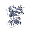 2rfdC 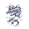 2rfeC 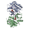 2gs7S C: citing same article ( S: Starting model for refinement |
|---|---|
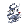
| Similar structure data |

-Links
 PDBj
PDBj








- Assembly
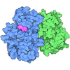
| Deposited unit | 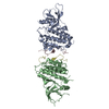
| ||||||||
|---|---|---|---|---|---|---|---|---|---|
| 1 | 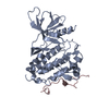
| ||||||||
| 2 | 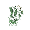
| ||||||||
| Unit cell |
|
-Components
| #1: Protein | Mass: 37563.457 Da / Num. of mol.: 2 / Fragment: Protein kinase domain Source method: isolated from a genetically manipulated source Source: (gene. exp.) Homo sapiens (human) / Gene: EGFR, ERBB1 / Plasmid: pFASTBAC-HT / Production host:   Spodoptera frugiperda (fall armyworm) / Strain (production host): Sf9 Spodoptera frugiperda (fall armyworm) / Strain (production host): Sf9References: UniProt: P00533, receptor protein-tyrosine kinase #2: Protein | Mass: 6866.682 Da / Num. of mol.: 2 / Fragment: sequence database residues, 315-374 Source method: isolated from a genetically manipulated source Source: (gene. exp.) Homo sapiens (human) / Gene: ERRFI1, MIG6 / Plasmid: pGEX6p1 / Species (production host): Escherichia coli / Production host:   Escherichia coli BL21(DE3) (bacteria) / Strain (production host): BL21(DE3) / References: UniProt: Q9UJM3 Escherichia coli BL21(DE3) (bacteria) / Strain (production host): BL21(DE3) / References: UniProt: Q9UJM3 |
|---|
-Experimental details
-Experiment
| Experiment | Method: X-RAY DIFFRACTION / Number of used crystals: 1 |
|---|
- Sample preparation
| Crystal | Density Matthews: 2.13 Å3/Da / Density % sol: 42.37 % |
|---|---|
| Crystal grow | Temperature: 293 K / Method: vapor diffusion, hanging drop / pH: 5 Details: 20% PEG3350, 300 mM KAC, 10% Glycerol and 100 mM NaAc, pH 5.0, vapor diffusion, hanging drop, temperature 293K |
-Data collection
| Diffraction | Mean temperature: 100 K |
|---|---|
| Diffraction source | Source: SYNCHROTRON / Site: ALS  / Beamline: 8.2.1 / Wavelength: 1 Å / Beamline: 8.2.1 / Wavelength: 1 Å |
| Detector | Type: ADSC QUANTUM 4 / Detector: CCD / Date: Jun 7, 2006 |
| Radiation | Protocol: SINGLE WAVELENGTH / Monochromatic (M) / Laue (L): M / Scattering type: x-ray |
| Radiation wavelength | Wavelength: 1 Å / Relative weight: 1 |
| Reflection | Resolution: 3.5→50 Å / Num. obs: 9081 / % possible obs: 91.8 % / Redundancy: 3.3 % / Rmerge(I) obs: 0.147 / Χ2: 1.049 / Net I/σ(I): 6.7 |
| Reflection shell | Resolution: 3.5→3.63 Å / Redundancy: 2.2 % / Rmerge(I) obs: 0.41 / Mean I/σ(I) obs: 1.8 / Num. unique all: 736 / Χ2: 0.935 / % possible all: 74.3 |
-Phasing
| Phasing | Method: molecular replacement |
|---|
- Processing
| Software |
| ||||||||||||||||||||||||||||||||
|---|---|---|---|---|---|---|---|---|---|---|---|---|---|---|---|---|---|---|---|---|---|---|---|---|---|---|---|---|---|---|---|---|---|
| Refinement | Method to determine structure: MOLECULAR REPLACEMENT Starting model: PDB entry 2GS7 Resolution: 3.5→47.11 Å / Isotropic thermal model: isotropic / Cross valid method: THROUGHOUT / σ(F): 0 / Stereochemistry target values: Engh & Huber
| ||||||||||||||||||||||||||||||||
| Solvent computation | Bsol: 67.047 Å2 | ||||||||||||||||||||||||||||||||
| Displacement parameters | Biso mean: 68.346 Å2
| ||||||||||||||||||||||||||||||||
| Refinement step | Cycle: LAST / Resolution: 3.5→47.11 Å
| ||||||||||||||||||||||||||||||||
| Refine LS restraints |
| ||||||||||||||||||||||||||||||||
| Xplor file | Serial no: 1 / Param file: protein_rep.param |
