- PDB-2reu: Crystal Structure of the C-terminal of Sau3AI fragment -
+
Open data
ID or keywords:
Loading...
-
Basic information
Entry
Database: PDB / ID: 2reu
Title
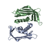
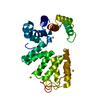
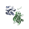
Crystal Structure of the C-terminal of Sau3AI fragment
Components
Type II restriction enzyme Sau3AI
Keywords
HYDROLASE / helix / beta / random coil / Endonuclease / Magnesium / Nuclease / Restriction system
Function / homology
Function and homology information
type II site-specific deoxyribonuclease / type II site-specific deoxyribonuclease activity / DNA restriction-modification system / DNA binding Similarity search - Function
DNA mismatch repair MutH/Type II restriction enzyme Sau3AI / DNA mismatch repair enzyme MutH / DNA mismatch repair enzyme MutH / DNA mismatch repair MutH/Type II restriction endonuclease superfamily / Restriction endonuclease type II-like Similarity search - Domain/homology
Av σ(I) over netI: 10 / Number: 302170 / Rmerge(I) obs: 0.071 / Χ2: 1.03 / D res high: 1.9 Å / D res low: 30 Å / Num. obs: 25678 / % possible obs: 96.4
Diffraction reflection shell
Highest resolution (Å)
Lowest resolution (Å)
% possible obs (%)
ID
Rmerge(I) obs
Chi squared
1.9
1.97
77.1
1
0.433
0.326
1.97
2.05
94.1
1
0.353
0.305
2.05
2.14
98.3
1
0.281
0.368
2.14
2.25
98.2
1
0.222
0.419
2.25
2.39
98.7
1
0.17
0.485
2.39
2.58
98.9
1
0.14
0.577
2.58
2.84
99.2
1
0.099
0.799
2.84
3.25
99.6
1
0.07
1.138
3.25
4.09
99.9
1
0.051
1.691
4.09
30
99.8
1
0.047
2.766
Reflection
Resolution: 1.9→30 Å / Num. obs: 25678 / % possible obs: 96.4 % / Rmerge(I) obs: 0.071 / Χ2: 1.034 / Net I/σ(I): 10
Reflection shell
Resolution (Å)
Rmerge(I) obs
Num. unique all
Χ2
Diffraction-ID
% possible all
1.9-1.97
0.433
2015
0.326
1
77.1
1.97-2.05
0.353
2473
0.305
1
94.1
2.05-2.14
0.281
2543
0.368
1
98.3
2.14-2.25
0.222
2580
0.419
1
98.2
2.25-2.39
0.17
2596
0.485
1
98.7
2.39-2.58
0.14
2609
0.577
1
98.9
2.58-2.84
0.099
2630
0.799
1
99.2
2.84-3.25
0.07
2659
1.138
1
99.6
3.25-4.09
0.051
2711
1.691
1
99.9
4.09-30
0.047
2862
2.766
1
99.8
-
Phasing
Phasing
Method: MAD
Phasing set
ID
1
2
3
Phasing MAD set
Clust-ID
Expt-ID
Set-ID
Wavelength (Å)
F double prime refined
F prime refined
1
3wavelength
1
0.9794
2.61
-10.43
1
3wavelength
2
0.9789
5.76
-5.41
1
3wavelength
3
0.9
3.67
-1.05
Phasing MAD set site
ID
Atom type symbol
B iso
Fract x
Fract y
Fract z
Occupancy
1
Se
21.372
0.247
0.175
0.23
1.072
2
Se
22.106
0.696
0.267
0.116
0.997
3
Se
21.997
0.676
0.202
0.254
0.839
4
Se
30.786
0.759
0.157
0.075
0.798
-
Processing
Software
Name
Version
Classification
NB
DENZO
datareduction
SCALEPACK
datascaling
SOLVE
2.13
phasing
REFMAC
refinement
PDB_EXTRACT
3
dataextraction
HKL-2000
datacollection
Refinement
Method to determine structure: MAD / Resolution: 1.9→28.35 Å / Cor.coef. Fo:Fc: 0.95 / Cor.coef. Fo:Fc free: 0.934 / SU B: 3.146 / SU ML: 0.095 / Cross valid method: THROUGHOUT / σ(F): 0 / ESU R: 0.157 / ESU R Free: 0.145 / Stereochemistry target values: MAXIMUM LIKELIHOOD / Details: HYDROGENS HAVE BEEN ADDED IN THE RIDING POSITIONS
Rfactor
Num. reflection
% reflection
Selection details
Rfree
0.236
1276
5 %
RANDOM
Rwork
0.199
-
-
-
obs
0.201
25634
96.4 %
-
Solvent computation
Ion probe radii: 0.8 Å / Shrinkage radii: 0.8 Å / VDW probe radii: 1.2 Å / Solvent model: MASK
Displacement parameters
Biso mean: 30.739 Å2
Baniso -1
Baniso -2
Baniso -3
1-
-1.15 Å2
0 Å2
0 Å2
2-
-
1.61 Å2
0 Å2
3-
-
-
-0.46 Å2
Refinement step
Cycle: LAST / Resolution: 1.9→28.35 Å
Protein
Nucleic acid
Ligand
Solvent
Total
Num. atoms
1961
0
1
225
2187
Refine LS restraints
Refine-ID
Type
Dev ideal
Dev ideal target
Number
X-RAY DIFFRACTION
r_bond_refined_d
0.008
0.022
2275
X-RAY DIFFRACTION
r_angle_refined_deg
1.031
1.938
3121
X-RAY DIFFRACTION
r_dihedral_angle_1_deg
4.883
5
307
X-RAY DIFFRACTION
r_dihedral_angle_2_deg
35.606
25.588
136
X-RAY DIFFRACTION
r_dihedral_angle_3_deg
13.929
15
448
X-RAY DIFFRACTION
r_dihedral_angle_4_deg
12.459
15
11
X-RAY DIFFRACTION
r_chiral_restr
0.08
0.2
316
X-RAY DIFFRACTION
r_gen_planes_refined
0.004
0.02
1815
X-RAY DIFFRACTION
r_nbd_refined
0.225
0.3
1023
X-RAY DIFFRACTION
r_nbtor_refined
0.321
0.5
1532
X-RAY DIFFRACTION
r_xyhbond_nbd_refined
0.231
0.5
328
X-RAY DIFFRACTION
r_symmetry_vdw_refined
0.156
0.3
23
X-RAY DIFFRACTION
r_symmetry_hbond_refined
0.256
0.5
28
X-RAY DIFFRACTION
r_mcbond_it
0.824
2
1331
X-RAY DIFFRACTION
r_mcangle_it
1.304
3
2134
X-RAY DIFFRACTION
r_scbond_it
0.712
2
1085
X-RAY DIFFRACTION
r_scangle_it
1.026
3
949
LS refinement shell
Resolution: 1.9→1.95 Å / Total num. of bins used: 20
Rfactor
Num. reflection
% reflection
Rfree
0.332
65
-
Rwork
0.294
1348
-
all
-
1413
-
obs
-
-
72.42 %
+
About Yorodumi
-
News
-
Feb 9, 2022. New format data for meta-information of EMDB entries
New format data for meta-information of EMDB entries
Version 3 of the EMDB header file is now the official format.
The previous official version 1.9 will be removed from the archive.
In the structure databanks used in Yorodumi, some data are registered as the other names, "COVID-19 virus" and "2019-nCoV". Here are the details of the virus and the list of structure data.
Jan 31, 2019. EMDB accession codes are about to change! (news from PDBe EMDB page)
EMDB accession codes are about to change! (news from PDBe EMDB page)
The allocation of 4 digits for EMDB accession codes will soon come to an end. Whilst these codes will remain in use, new EMDB accession codes will include an additional digit and will expand incrementally as the available range of codes is exhausted. The current 4-digit format prefixed with “EMD-” (i.e. EMD-XXXX) will advance to a 5-digit format (i.e. EMD-XXXXX), and so on. It is currently estimated that the 4-digit codes will be depleted around Spring 2019, at which point the 5-digit format will come into force.
The EM Navigator/Yorodumi systems omit the EMD- prefix.
Related info.:Q: What is EMD? / ID/Accession-code notation in Yorodumi/EM Navigator
Yorodumi is a browser for structure data from EMDB, PDB, SASBDB, etc.
This page is also the successor to EM Navigator detail page, and also detail information page/front-end page for Omokage search.
The word "yorodu" (or yorozu) is an old Japanese word meaning "ten thousand". "mi" (miru) is to see.
Related info.:EMDB / PDB / SASBDB / Comparison of 3 databanks / Yorodumi Search / Aug 31, 2016. New EM Navigator & Yorodumi / Yorodumi Papers / Jmol/JSmol / Function and homology information / Changes in new EM Navigator and Yorodumi
 Movie
Movie Controller


 Open data
 Basic information
 Components
 Keywords
 Function and homology information
 Staphylococcus aureus (bacteria)
 X-RAY DIFFRACTION /
  Authors
 Citation
 Structure visualization
 Downloads & links
 Other downloads
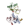


 PDBj
PDBj


 Assembly

 Mass: 24.305 Da / Num. of mol.: 1 / Source method: obtained synthetically / Formula: Mg
 Mass: 18.015 Da / Num. of mol.: 225 / Source method: isolated from a natural source / Formula: H2O
 Sample preparation
 / Beamline: 3W1A / Wavelength: 0.9789, 0.97945, 0.9
 Processing